Mercury »
PDB 1czs-1g4o »
1e7z »
Mercury in PDB 1e7z: Crystal Structure of the EMAP2/Rna Binding Domain of the P43 Protein From Human Aminoacyl-Trna Synthetase Complex
Protein crystallography data
The structure of Crystal Structure of the EMAP2/Rna Binding Domain of the P43 Protein From Human Aminoacyl-Trna Synthetase Complex, PDB code: 1e7z
was solved by
S.Pasqualato,
P.Kerjan,
L.Renault,
J.Menetrey,
M.Mirande,
J.Cherfils,
with X-Ray Crystallography technique. A brief refinement statistics is given in the table below:
| Resolution Low / High (Å) | 28.48 / 2.05 |
| Space group | P 1 21 1 |
| Cell size a, b, c (Å), α, β, γ (°) | 36.141, 53.938, 41.371, 90.00, 111.92, 90.00 |
| R / Rfree (%) | 22.1 / 27.2 |
Mercury Binding Sites:
The binding sites of Mercury atom in the Crystal Structure of the EMAP2/Rna Binding Domain of the P43 Protein From Human Aminoacyl-Trna Synthetase Complex
(pdb code 1e7z). This binding sites where shown within
5.0 Angstroms radius around Mercury atom.
In total 2 binding sites of Mercury where determined in the Crystal Structure of the EMAP2/Rna Binding Domain of the P43 Protein From Human Aminoacyl-Trna Synthetase Complex, PDB code: 1e7z:
Jump to Mercury binding site number: 1; 2;
In total 2 binding sites of Mercury where determined in the Crystal Structure of the EMAP2/Rna Binding Domain of the P43 Protein From Human Aminoacyl-Trna Synthetase Complex, PDB code: 1e7z:
Jump to Mercury binding site number: 1; 2;
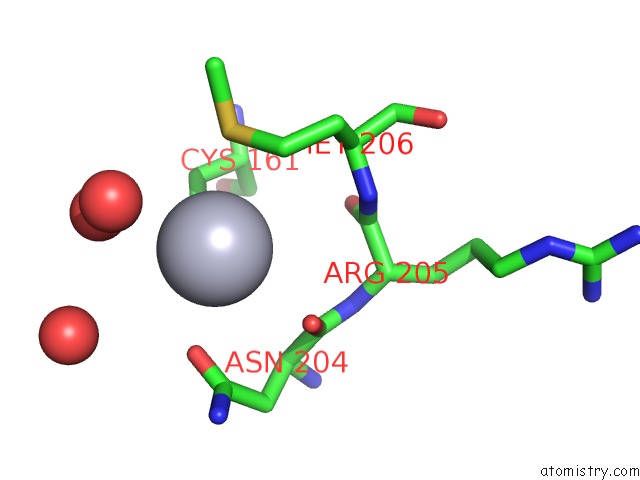
Mercury binding site 1 out of 2 in 1e7z
Go back to
Mercury binding site 1 out
of 2 in the Crystal Structure of the EMAP2/Rna Binding Domain of the P43 Protein From Human Aminoacyl-Trna Synthetase Complex
Mono view
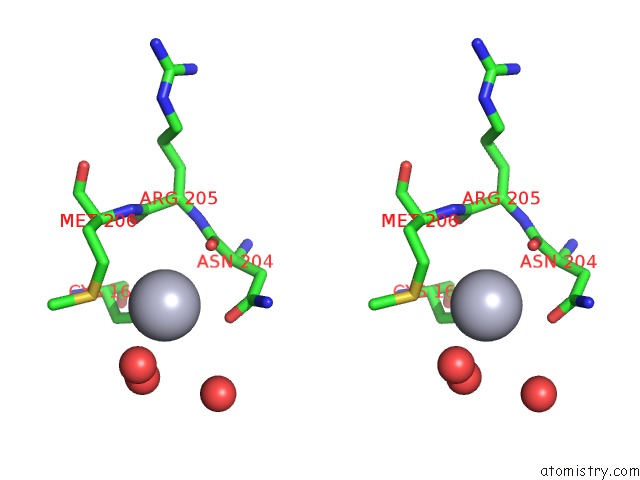
Stereo pair view
Mono view
Stereo pair view
A full contact list of Mercury with other atoms in the Hg binding
site number 1 of Crystal Structure of the EMAP2/Rna Binding Domain of the P43 Protein From Human Aminoacyl-Trna Synthetase Complex within 5.0Å range:
|
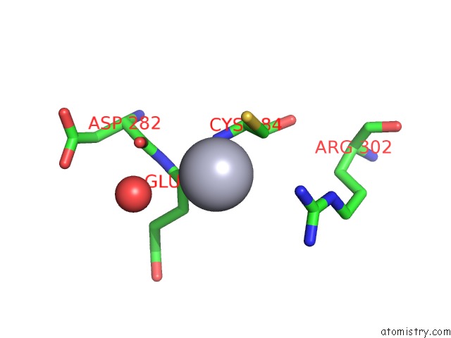
Mercury binding site 2 out of 2 in 1e7z
Go back to
Mercury binding site 2 out
of 2 in the Crystal Structure of the EMAP2/Rna Binding Domain of the P43 Protein From Human Aminoacyl-Trna Synthetase Complex
Mono view
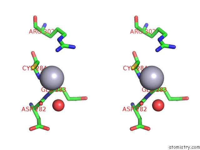
Stereo pair view
Mono view
Stereo pair view
A full contact list of Mercury with other atoms in the Hg binding
site number 2 of Crystal Structure of the EMAP2/Rna Binding Domain of the P43 Protein From Human Aminoacyl-Trna Synthetase Complex within 5.0Å range:
|
Reference:
L.Renault,
P.Kerjan,
S.Pasqualato,
J.Menetrey,
J.C.Robinson,
S.Kawaguchi,
D.G.Vassylyev,
S.Yokoyama,
M.Mirande,
J.Cherfils.
Structure of the Emapii Domain of Human Aminoacyl- Trna Synthetase Complex Reveals Evolutionary Dimeric Mimicry Embo J. V. 20 570 2001.
ISSN: ISSN 0261-4189
PubMed: 11157763
DOI: 10.1093/EMBOJ/20.3.570
Page generated: Sat Aug 10 23:38:00 2024
ISSN: ISSN 0261-4189
PubMed: 11157763
DOI: 10.1093/EMBOJ/20.3.570
Last articles
Fe in 7LZKFe in 7LYX
Fe in 7LXL
Fe in 7LW8
Fe in 7LWA
Fe in 7LW9
Fe in 7LJU
Fe in 7LJT
Fe in 7LJS
Fe in 7LW7