Mercury »
PDB 1rsv-1yp2 »
1t3s »
Mercury in PDB 1t3s: Structural Analysis of the Voltage-Dependent Calcium Channel Beta Subunit Functional Core
Protein crystallography data
The structure of Structural Analysis of the Voltage-Dependent Calcium Channel Beta Subunit Functional Core, PDB code: 1t3s
was solved by
Y.Opatowsky,
C.-C.Chen,
K.P.Campbell,
J.A.Hirsch,
with X-Ray Crystallography technique. A brief refinement statistics is given in the table below:
| Resolution Low / High (Å) | 81.65 / 2.30 |
| Space group | P 21 21 2 |
| Cell size a, b, c (Å), α, β, γ (°) | 74.066, 163.841, 34.761, 90.00, 90.00, 90.00 |
| R / Rfree (%) | 26.1 / 27.7 |
Mercury Binding Sites:
The binding sites of Mercury atom in the Structural Analysis of the Voltage-Dependent Calcium Channel Beta Subunit Functional Core
(pdb code 1t3s). This binding sites where shown within
5.0 Angstroms radius around Mercury atom.
In total only one binding site of Mercury was determined in the Structural Analysis of the Voltage-Dependent Calcium Channel Beta Subunit Functional Core, PDB code: 1t3s:
In total only one binding site of Mercury was determined in the Structural Analysis of the Voltage-Dependent Calcium Channel Beta Subunit Functional Core, PDB code: 1t3s:
Mercury binding site 1 out of 1 in 1t3s
Go back to
Mercury binding site 1 out
of 1 in the Structural Analysis of the Voltage-Dependent Calcium Channel Beta Subunit Functional Core
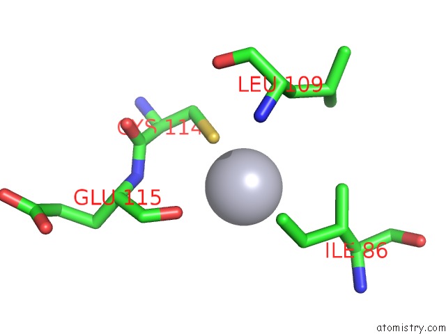
Mono view
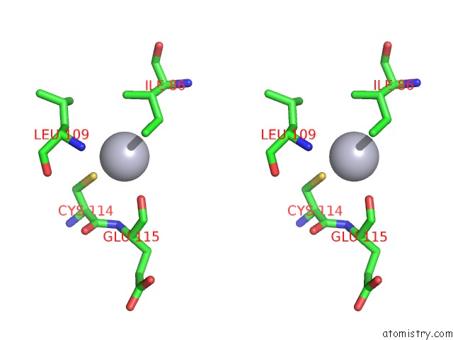
Stereo pair view
Mono view
Stereo pair view
A full contact list of Mercury with other atoms in the Hg binding
site number 1 of Structural Analysis of the Voltage-Dependent Calcium Channel Beta Subunit Functional Core within 5.0Å range:
|
Reference:
Y.Opatowsky,
C.C.Chen,
K.P.Campbell,
J.A.Hirsch.
Structural Analysis of the Voltage-Dependent Calcium Channel Beta Subunit Functional Core and Its Complex with the Alpha 1 Interaction Domain. Neuron V. 42 387 2004.
ISSN: ISSN 0896-6273
PubMed: 15134636
DOI: 10.1093/HMG/DDH162
Page generated: Fri Aug 8 09:33:11 2025
ISSN: ISSN 0896-6273
PubMed: 15134636
DOI: 10.1093/HMG/DDH162
Last articles
Mn in 8JYHMn in 8JQE
Mn in 8JQ6
Mn in 8JQ4
Mn in 8JQ3
Mn in 8JKK
Mn in 8JNK
Mn in 8JNR
Mn in 8IRI
Mn in 8JGP