Mercury »
PDB 1yu1-2epm »
2di4 »
Mercury in PDB 2di4: Crystal Structure of the Ftsh Protease Domain
Protein crystallography data
The structure of Crystal Structure of the Ftsh Protease Domain, PDB code: 2di4
was solved by
R.Suno,
H.Niwa,
D.Tsuchiya,
X.Zhang,
M.Yoshida,
K.Morikawa,
with X-Ray Crystallography technique. A brief refinement statistics is given in the table below:
| Resolution Low / High (Å) | 20.00 / 2.79 |
| Space group | P 63 |
| Cell size a, b, c (Å), α, β, γ (°) | 116.800, 116.800, 63.500, 90.00, 90.00, 120.00 |
| R / Rfree (%) | 25.1 / 29.9 |
Mercury Binding Sites:
The binding sites of Mercury atom in the Crystal Structure of the Ftsh Protease Domain
(pdb code 2di4). This binding sites where shown within
5.0 Angstroms radius around Mercury atom.
In total 2 binding sites of Mercury where determined in the Crystal Structure of the Ftsh Protease Domain, PDB code: 2di4:
Jump to Mercury binding site number: 1; 2;
In total 2 binding sites of Mercury where determined in the Crystal Structure of the Ftsh Protease Domain, PDB code: 2di4:
Jump to Mercury binding site number: 1; 2;
Mercury binding site 1 out of 2 in 2di4
Go back to
Mercury binding site 1 out
of 2 in the Crystal Structure of the Ftsh Protease Domain
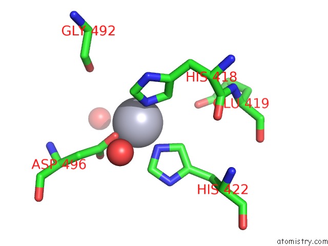
Mono view
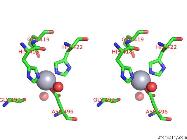
Stereo pair view
Mono view
Stereo pair view
A full contact list of Mercury with other atoms in the Hg binding
site number 1 of Crystal Structure of the Ftsh Protease Domain within 5.0Å range:
|
Mercury binding site 2 out of 2 in 2di4
Go back to
Mercury binding site 2 out
of 2 in the Crystal Structure of the Ftsh Protease Domain
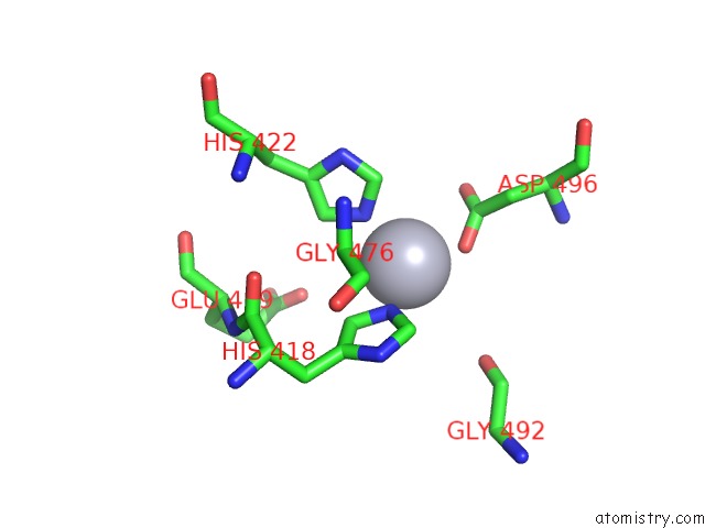
Mono view
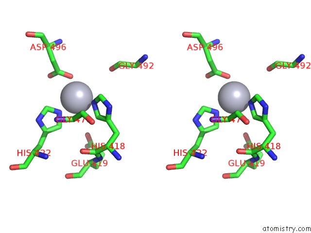
Stereo pair view
Mono view
Stereo pair view
A full contact list of Mercury with other atoms in the Hg binding
site number 2 of Crystal Structure of the Ftsh Protease Domain within 5.0Å range:
|
Reference:
R.Suno,
H.Niwa,
D.Tsuchiya,
X.Zhang,
M.Yoshida,
K.Morikawa.
Structure of the Whole Cytosolic Region of Atp-Dependent Protease Ftsh Mol.Cell V. 22 575 2006.
ISSN: ISSN 1097-2765
PubMed: 16762831
DOI: 10.1016/J.MOLCEL.2006.04.020
Page generated: Fri Aug 8 09:47:37 2025
ISSN: ISSN 1097-2765
PubMed: 16762831
DOI: 10.1016/J.MOLCEL.2006.04.020
Last articles
Na in 1S0ANa in 1S1K
Na in 1S09
Na in 1RZT
Na in 1S08
Na in 1S07
Na in 1S06
Na in 1RYS
Na in 1RWT
Na in 1RWH