Mercury »
PDB 2esw-2o1f »
2ga8 »
Mercury in PDB 2ga8: Crystal Structure of YFH7 From Saccharomyces Cerevisiae: A Putative P-Loop Containing Kinase with A Circular Permutation.
Protein crystallography data
The structure of Crystal Structure of YFH7 From Saccharomyces Cerevisiae: A Putative P-Loop Containing Kinase with A Circular Permutation., PDB code: 2ga8
was solved by
V.Chaptal,
S.Morera,
with X-Ray Crystallography technique. A brief refinement statistics is given in the table below:
| Resolution Low / High (Å) | 20.00 / 1.77 |
| Space group | H 3 |
| Cell size a, b, c (Å), α, β, γ (°) | 135.278, 135.278, 48.476, 90.00, 90.00, 120.00 |
| R / Rfree (%) | 19.4 / 21.9 |
Mercury Binding Sites:
The binding sites of Mercury atom in the Crystal Structure of YFH7 From Saccharomyces Cerevisiae: A Putative P-Loop Containing Kinase with A Circular Permutation.
(pdb code 2ga8). This binding sites where shown within
5.0 Angstroms radius around Mercury atom.
In total 4 binding sites of Mercury where determined in the Crystal Structure of YFH7 From Saccharomyces Cerevisiae: A Putative P-Loop Containing Kinase with A Circular Permutation., PDB code: 2ga8:
Jump to Mercury binding site number: 1; 2; 3; 4;
In total 4 binding sites of Mercury where determined in the Crystal Structure of YFH7 From Saccharomyces Cerevisiae: A Putative P-Loop Containing Kinase with A Circular Permutation., PDB code: 2ga8:
Jump to Mercury binding site number: 1; 2; 3; 4;
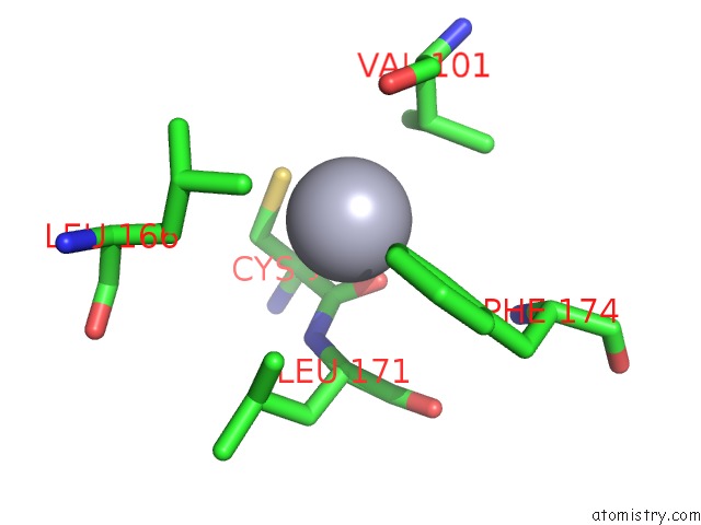
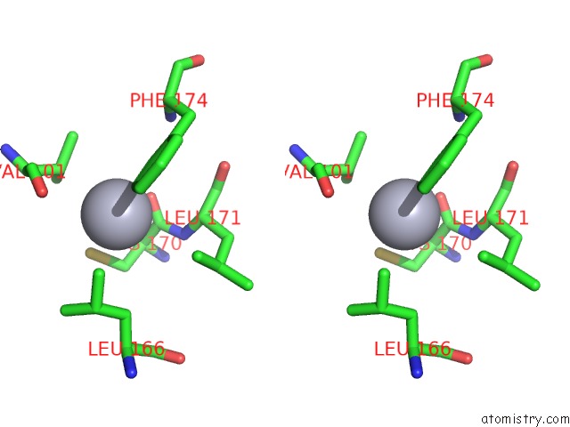
Mercury binding site 1 out of 4 in 2ga8
Go back to
Mercury binding site 1 out
of 4 in the Crystal Structure of YFH7 From Saccharomyces Cerevisiae: A Putative P-Loop Containing Kinase with A Circular Permutation.
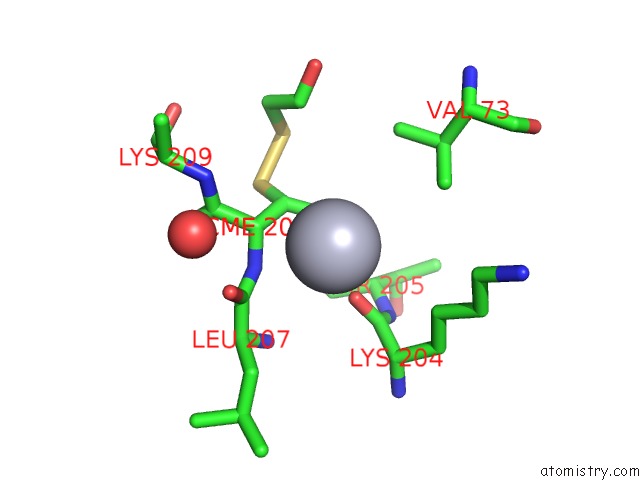
Mono view
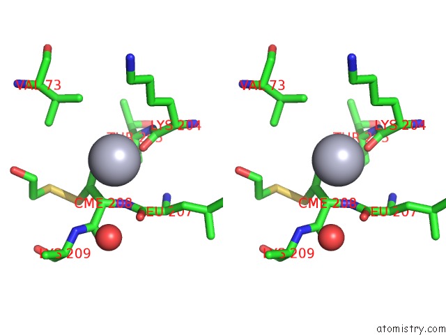
Stereo pair view
Mono view
Stereo pair view
A full contact list of Mercury with other atoms in the Hg binding
site number 1 of Crystal Structure of YFH7 From Saccharomyces Cerevisiae: A Putative P-Loop Containing Kinase with A Circular Permutation. within 5.0Å range:
|
Mercury binding site 2 out of 4 in 2ga8
Go back to
Mercury binding site 2 out
of 4 in the Crystal Structure of YFH7 From Saccharomyces Cerevisiae: A Putative P-Loop Containing Kinase with A Circular Permutation.
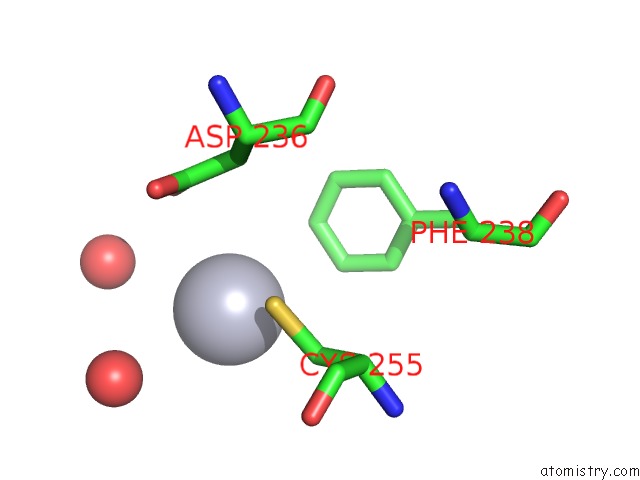
Mono view
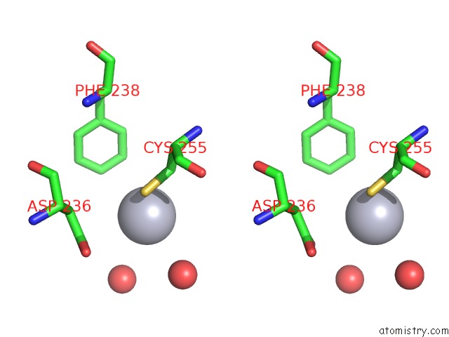
Stereo pair view
Mono view
Stereo pair view
A full contact list of Mercury with other atoms in the Hg binding
site number 2 of Crystal Structure of YFH7 From Saccharomyces Cerevisiae: A Putative P-Loop Containing Kinase with A Circular Permutation. within 5.0Å range:
|
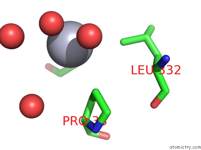
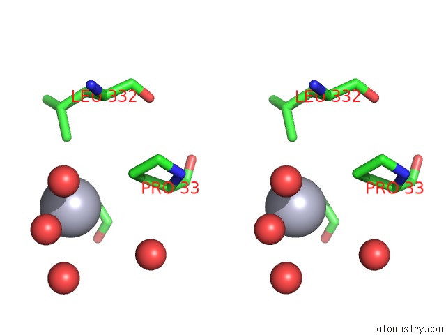
Mercury binding site 3 out of 4 in 2ga8
Go back to
Mercury binding site 3 out
of 4 in the Crystal Structure of YFH7 From Saccharomyces Cerevisiae: A Putative P-Loop Containing Kinase with A Circular Permutation.
Mono view
Stereo pair view
Mono view
Stereo pair view
A full contact list of Mercury with other atoms in the Hg binding
site number 3 of Crystal Structure of YFH7 From Saccharomyces Cerevisiae: A Putative P-Loop Containing Kinase with A Circular Permutation. within 5.0Å range:
|
Mercury binding site 4 out of 4 in 2ga8
Go back to
Mercury binding site 4 out
of 4 in the Crystal Structure of YFH7 From Saccharomyces Cerevisiae: A Putative P-Loop Containing Kinase with A Circular Permutation.
Mono view
Stereo pair view
Mono view
Stereo pair view
A full contact list of Mercury with other atoms in the Hg binding
site number 4 of Crystal Structure of YFH7 From Saccharomyces Cerevisiae: A Putative P-Loop Containing Kinase with A Circular Permutation. within 5.0Å range:
|
Reference:
V.Gueguen-Chaignon,
V.Chaptal,
L.Lariviere,
N.Costa,
P.Lopes,
S.Morera,
S.Nessler.
Crystal Structure and Functional Analysis Identify the P-Loop Containing Protein YFH7 of Saccharomyces Cerevisiae As An Atp-Dependent Kinase. Proteins V. 71 804 2007.
ISSN: ISSN 0887-3585
PubMed: 18004758
DOI: 10.1002/PROT.21740
Page generated: Fri Aug 8 09:49:21 2025
ISSN: ISSN 0887-3585
PubMed: 18004758
DOI: 10.1002/PROT.21740
Last articles
Mn in 9L56Mn in 9KOQ
Mn in 9KOJ
Mn in 9KQC
Mn in 9JOA
Mn in 9JOB
Mn in 9JHN
Mn in 9JL7
Mn in 9JHL
Mn in 9JH9