Mercury »
PDB 2o1g-3b4f »
357d »
Mercury in PDB 357d: 3.5 A Structure of Fragment I From E. Coli 5S Rrna
Protein crystallography data
The structure of 3.5 A Structure of Fragment I From E. Coli 5S Rrna, PDB code: 357d
was solved by
C.C.Correll,
B.Freeborn,
P.B.Moore,
T.A.Steitz,
with X-Ray Crystallography technique. A brief refinement statistics is given in the table below:
| Resolution Low / High (Å) | 10.00 / 3.50 |
| Space group | P 61 2 2 |
| Cell size a, b, c (Å), α, β, γ (°) | 58.670, 58.670, 248.840, 90.00, 90.00, 120.00 |
| R / Rfree (%) | 31.2 / 26.5 |
Other elements in 357d:
The structure of 3.5 A Structure of Fragment I From E. Coli 5S Rrna also contains other interesting chemical elements:
| Magnesium | (Mg) | 8 atoms |
Mercury Binding Sites:
The binding sites of Mercury atom in the 3.5 A Structure of Fragment I From E. Coli 5S Rrna
(pdb code 357d). This binding sites where shown within
5.0 Angstroms radius around Mercury atom.
In total only one binding site of Mercury was determined in the 3.5 A Structure of Fragment I From E. Coli 5S Rrna, PDB code: 357d:
In total only one binding site of Mercury was determined in the 3.5 A Structure of Fragment I From E. Coli 5S Rrna, PDB code: 357d:
Mercury binding site 1 out of 1 in 357d
Go back to
Mercury binding site 1 out
of 1 in the 3.5 A Structure of Fragment I From E. Coli 5S Rrna
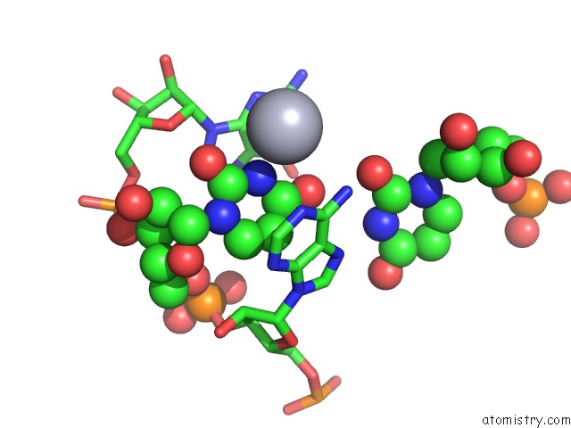
Mono view
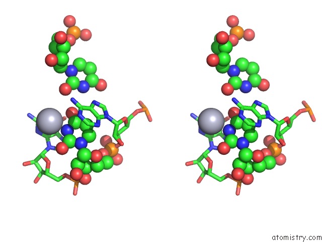
Stereo pair view
Mono view
Stereo pair view
A full contact list of Mercury with other atoms in the Hg binding
site number 1 of 3.5 A Structure of Fragment I From E. Coli 5S Rrna within 5.0Å range:
|
Reference:
C.C.Correll,
B.Freeborn,
P.B.Moore,
T.A.Steitz.
Metals, Motifs, and Recognition in the Crystal Structure of A 5S Rrna Domain. Cell(Cambridge,Mass.) V. 91 705 1997.
ISSN: ISSN 0092-8674
PubMed: 9393863
DOI: 10.1016/S0092-8674(00)80457-2
Page generated: Sun Aug 11 03:09:05 2024
ISSN: ISSN 0092-8674
PubMed: 9393863
DOI: 10.1016/S0092-8674(00)80457-2
Last articles
Fe in 7M1KFe in 7M1J
Fe in 7M1I
Fe in 7M0H
Fe in 7M0F
Fe in 7LZO
Fe in 7LXD
Fe in 7LZN
Fe in 7LZK
Fe in 7LYX