Mercury »
PDB 3kbc-3wa8 »
3kp9 »
Mercury in PDB 3kp9: Structure of A Bacterial Homolog of Vitamin K Epoxide Reductase
Enzymatic activity of Structure of A Bacterial Homolog of Vitamin K Epoxide Reductase
All present enzymatic activity of Structure of A Bacterial Homolog of Vitamin K Epoxide Reductase:
1.1.4.2;
1.1.4.2;
Protein crystallography data
The structure of Structure of A Bacterial Homolog of Vitamin K Epoxide Reductase, PDB code: 3kp9
was solved by
W.Li,
S.Schulman,
R.J.Dutton,
D.Boyd,
J.Beckwith,
T.A.Rapoport,
with X-Ray Crystallography technique. A brief refinement statistics is given in the table below:
| Resolution Low / High (Å) | 44.83 / 3.60 |
| Space group | P 61 |
| Cell size a, b, c (Å), α, β, γ (°) | 136.942, 136.942, 68.480, 90.00, 90.00, 120.00 |
| R / Rfree (%) | 25.1 / 30.5 |
Mercury Binding Sites:
The binding sites of Mercury atom in the Structure of A Bacterial Homolog of Vitamin K Epoxide Reductase
(pdb code 3kp9). This binding sites where shown within
5.0 Angstroms radius around Mercury atom.
In total 2 binding sites of Mercury where determined in the Structure of A Bacterial Homolog of Vitamin K Epoxide Reductase, PDB code: 3kp9:
Jump to Mercury binding site number: 1; 2;
In total 2 binding sites of Mercury where determined in the Structure of A Bacterial Homolog of Vitamin K Epoxide Reductase, PDB code: 3kp9:
Jump to Mercury binding site number: 1; 2;
Mercury binding site 1 out of 2 in 3kp9
Go back to
Mercury binding site 1 out
of 2 in the Structure of A Bacterial Homolog of Vitamin K Epoxide Reductase
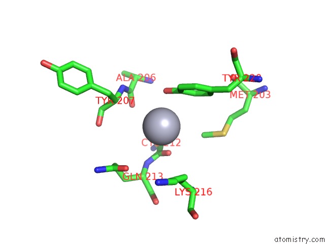
Mono view
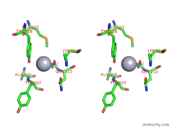
Stereo pair view
Mono view
Stereo pair view
A full contact list of Mercury with other atoms in the Hg binding
site number 1 of Structure of A Bacterial Homolog of Vitamin K Epoxide Reductase within 5.0Å range:
|
Mercury binding site 2 out of 2 in 3kp9
Go back to
Mercury binding site 2 out
of 2 in the Structure of A Bacterial Homolog of Vitamin K Epoxide Reductase
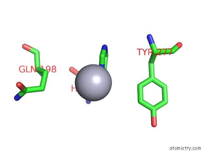
Mono view
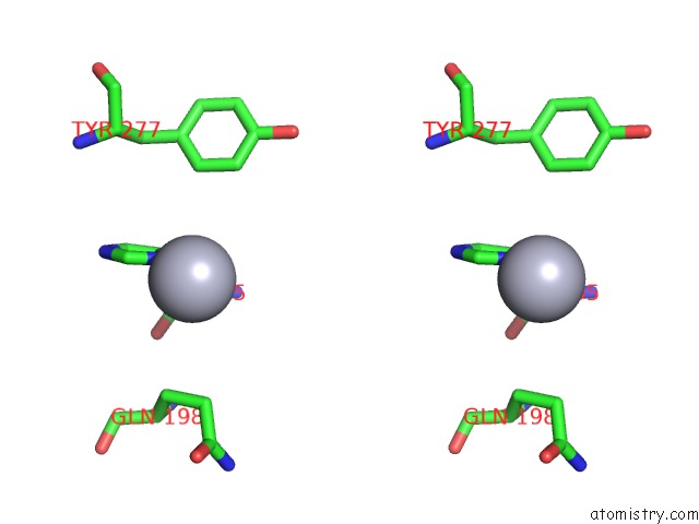
Stereo pair view
Mono view
Stereo pair view
A full contact list of Mercury with other atoms in the Hg binding
site number 2 of Structure of A Bacterial Homolog of Vitamin K Epoxide Reductase within 5.0Å range:
|
Reference:
W.Li,
S.Schulman,
R.J.Dutton,
D.Boyd,
J.Beckwith,
T.A.Rapoport.
Structure of A Bacterial Homologue of Vitamin K Epoxide Reductase. Nature V. 463 507 2010.
ISSN: ISSN 0028-0836
PubMed: 20110994
DOI: 10.1038/NATURE08720
Page generated: Fri Aug 8 10:12:29 2025
ISSN: ISSN 0028-0836
PubMed: 20110994
DOI: 10.1038/NATURE08720
Last articles
Mn in 3NIOMn in 3NWK
Mn in 3O1R
Mn in 3NVT
Mn in 3O1P
Mn in 3O1O
Mn in 3O1M
Mn in 3NV8
Mn in 3NUE
Mn in 3NQB