Mercury »
PDB 3kbc-3wa8 »
3ldi »
Mercury in PDB 3ldi: Crystal Structure of Aprotinin in Complex with Sucrose Octasulfate: Unusual Interactions and Implication For Heparin Binding
Protein crystallography data
The structure of Crystal Structure of Aprotinin in Complex with Sucrose Octasulfate: Unusual Interactions and Implication For Heparin Binding, PDB code: 3ldi
was solved by
I.S.Yang,
T.G.Kim,
B.S.Park,
K.H.Kim,
with X-Ray Crystallography technique. A brief refinement statistics is given in the table below:
| Resolution Low / High (Å) | 34.77 / 2.20 |
| Space group | P 63 2 2 |
| Cell size a, b, c (Å), α, β, γ (°) | 119.080, 119.080, 110.794, 90.00, 90.00, 120.00 |
| R / Rfree (%) | 20.6 / 23.4 |
Mercury Binding Sites:
The binding sites of Mercury atom in the Crystal Structure of Aprotinin in Complex with Sucrose Octasulfate: Unusual Interactions and Implication For Heparin Binding
(pdb code 3ldi). This binding sites where shown within
5.0 Angstroms radius around Mercury atom.
In total 5 binding sites of Mercury where determined in the Crystal Structure of Aprotinin in Complex with Sucrose Octasulfate: Unusual Interactions and Implication For Heparin Binding, PDB code: 3ldi:
Jump to Mercury binding site number: 1; 2; 3; 4; 5;
In total 5 binding sites of Mercury where determined in the Crystal Structure of Aprotinin in Complex with Sucrose Octasulfate: Unusual Interactions and Implication For Heparin Binding, PDB code: 3ldi:
Jump to Mercury binding site number: 1; 2; 3; 4; 5;
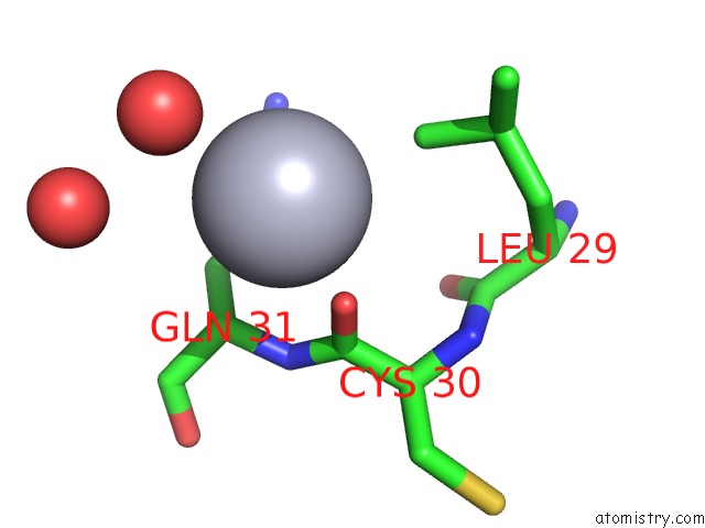
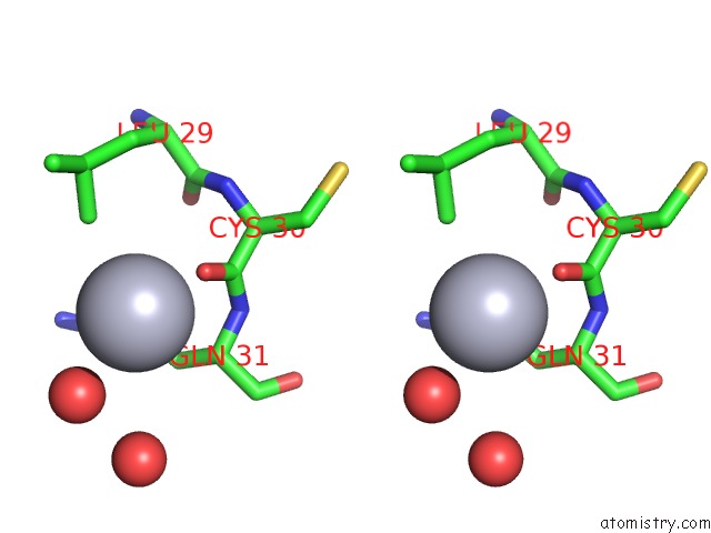
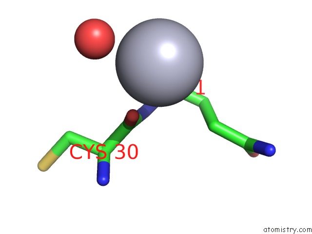
Mercury binding site 1 out of 5 in 3ldi
Go back to
Mercury binding site 1 out
of 5 in the Crystal Structure of Aprotinin in Complex with Sucrose Octasulfate: Unusual Interactions and Implication For Heparin Binding
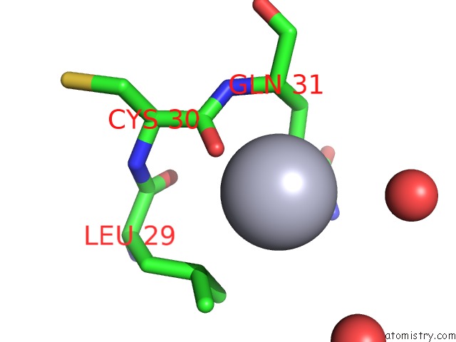
Mono view
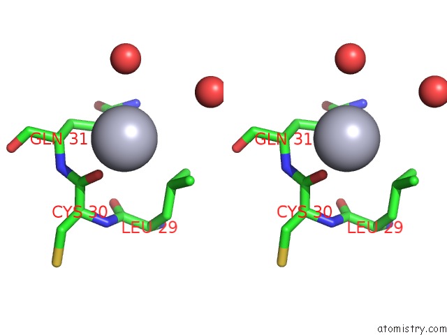
Stereo pair view
Mono view
Stereo pair view
|
|
A full contact list of Mercury with other atoms in the Hg binding
site number 1 of Crystal Structure of Aprotinin in Complex with Sucrose Octasulfate: Unusual Interactions and Implication For Heparin Binding within 5.0Å range:
|
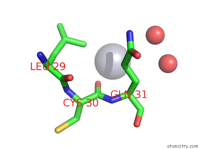
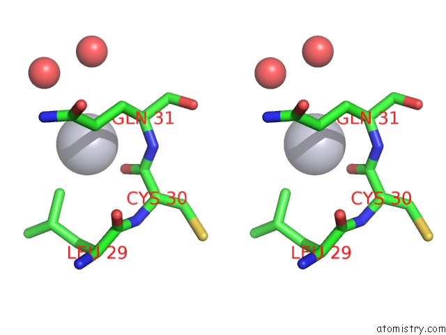
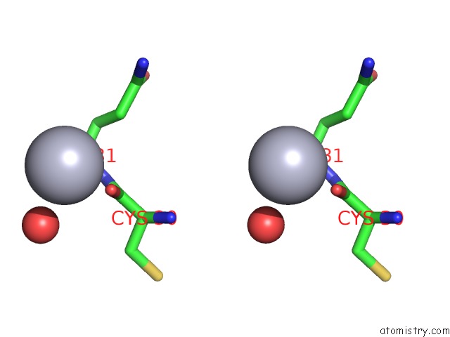
Mercury binding site 2 out of 5 in 3ldi
Go back to
Mercury binding site 2 out
of 5 in the Crystal Structure of Aprotinin in Complex with Sucrose Octasulfate: Unusual Interactions and Implication For Heparin Binding
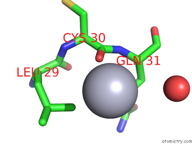
Mono view
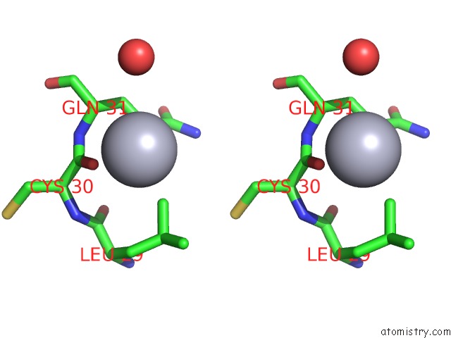
Stereo pair view
Mono view
Stereo pair view
|
|
A full contact list of Mercury with other atoms in the Hg binding
site number 2 of Crystal Structure of Aprotinin in Complex with Sucrose Octasulfate: Unusual Interactions and Implication For Heparin Binding within 5.0Å range:
|
Mercury binding site 3 out of 5 in 3ldi
Go back to
Mercury binding site 3 out
of 5 in the Crystal Structure of Aprotinin in Complex with Sucrose Octasulfate: Unusual Interactions and Implication For Heparin Binding
Mono view
Stereo pair view
Mono view
Stereo pair view
|
|
A full contact list of Mercury with other atoms in the Hg binding
site number 3 of Crystal Structure of Aprotinin in Complex with Sucrose Octasulfate: Unusual Interactions and Implication For Heparin Binding within 5.0Å range:
|
Mercury binding site 4 out of 5 in 3ldi
Go back to
Mercury binding site 4 out
of 5 in the Crystal Structure of Aprotinin in Complex with Sucrose Octasulfate: Unusual Interactions and Implication For Heparin Binding
Mono view
Stereo pair view
Mono view
Stereo pair view
|
|
A full contact list of Mercury with other atoms in the Hg binding
site number 4 of Crystal Structure of Aprotinin in Complex with Sucrose Octasulfate: Unusual Interactions and Implication For Heparin Binding within 5.0Å range:
|
Mercury binding site 5 out of 5 in 3ldi
Go back to
Mercury binding site 5 out
of 5 in the Crystal Structure of Aprotinin in Complex with Sucrose Octasulfate: Unusual Interactions and Implication For Heparin Binding
Mono view
Stereo pair view
Mono view
Stereo pair view
|
|
A full contact list of Mercury with other atoms in the Hg binding
site number 5 of Crystal Structure of Aprotinin in Complex with Sucrose Octasulfate: Unusual Interactions and Implication For Heparin Binding within 5.0Å range:
|
Reference:
I.S.Yang,
T.G.Kim,
B.S.Park,
K.J.Cho,
J.-H.Lee,
Y.Park,
K.H.Kim.
Crystal Structures of Aprotinin and Its Complex with Sucrose Octasulfate Reveal Multiple Modes of Interactions with Implications For Heparin Binding Biochem.Biophys.Res.Commun. V. 397 429 2010.
ISSN: ISSN 0006-291X
PubMed: 20529698
DOI: 10.1016/J.BBRC.2010.05.113
Page generated: Sun Aug 11 04:00:31 2024
ISSN: ISSN 0006-291X
PubMed: 20529698
DOI: 10.1016/J.BBRC.2010.05.113
Last articles
Zn in 9MJ5Zn in 9HNW
Zn in 9G0L
Zn in 9FNE
Zn in 9DZN
Zn in 9E0I
Zn in 9D32
Zn in 9DAK
Zn in 8ZXC
Zn in 8ZUF