Mercury »
PDB 3kbc-3wa8 »
3nj9 »
Mercury in PDB 3nj9: Crystal Structure of Carbonic Anhydrase II in Complex with A Nir Inhibitor
Enzymatic activity of Crystal Structure of Carbonic Anhydrase II in Complex with A Nir Inhibitor
All present enzymatic activity of Crystal Structure of Carbonic Anhydrase II in Complex with A Nir Inhibitor:
4.2.1.1;
4.2.1.1;
Protein crystallography data
The structure of Crystal Structure of Carbonic Anhydrase II in Complex with A Nir Inhibitor, PDB code: 3nj9
was solved by
C.Temperini,
A.Cecchi,
with X-Ray Crystallography technique. A brief refinement statistics is given in the table below:
| Resolution Low / High (Å) | 10.55 / 2.00 |
| Space group | P 1 21 1 |
| Cell size a, b, c (Å), α, β, γ (°) | 42.070, 41.540, 72.390, 90.00, 104.45, 90.00 |
| R / Rfree (%) | 20 / 25.7 |
Other elements in 3nj9:
The structure of Crystal Structure of Carbonic Anhydrase II in Complex with A Nir Inhibitor also contains other interesting chemical elements:
| Zinc | (Zn) | 1 atom |
Mercury Binding Sites:
The binding sites of Mercury atom in the Crystal Structure of Carbonic Anhydrase II in Complex with A Nir Inhibitor
(pdb code 3nj9). This binding sites where shown within
5.0 Angstroms radius around Mercury atom.
In total only one binding site of Mercury was determined in the Crystal Structure of Carbonic Anhydrase II in Complex with A Nir Inhibitor, PDB code: 3nj9:
In total only one binding site of Mercury was determined in the Crystal Structure of Carbonic Anhydrase II in Complex with A Nir Inhibitor, PDB code: 3nj9:
Mercury binding site 1 out of 1 in 3nj9
Go back to
Mercury binding site 1 out
of 1 in the Crystal Structure of Carbonic Anhydrase II in Complex with A Nir Inhibitor
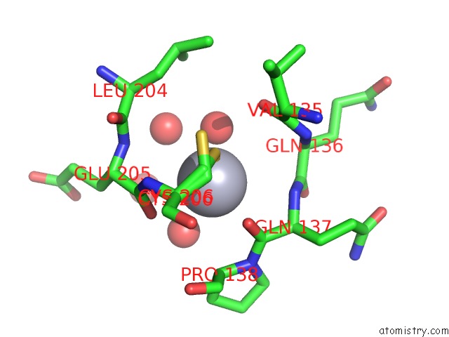
Mono view
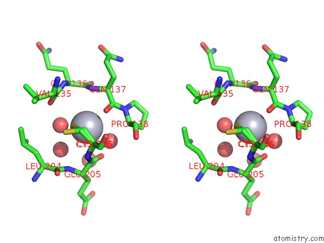
Stereo pair view
Mono view
Stereo pair view
|
|
A full contact list of Mercury with other atoms in the Hg binding
site number 1 of Crystal Structure of Carbonic Anhydrase II in Complex with A Nir Inhibitor within 5.0Å range:
|
Reference:
C.Temperini,
A.Cecchi.
Crystal Structure of Carbonic Anhydrase II in Complex with A Nir Inhibitor To Be Published.
Page generated: Sun Aug 11 04:06:07 2024
Last articles
Ca in 3U3WCa in 3U27
Ca in 3U24
Ca in 3U1W
Ca in 3U17
Ca in 3U16
Ca in 3U0K
Ca in 3TZ1
Ca in 3TVC
Ca in 3TTO