Mercury »
PDB 3kbc-3wa8 »
3oxh »
Mercury in PDB 3oxh: Mycobacterium Tuberculosis Kinase Inhibitor Homolog RV0577
Protein crystallography data
The structure of Mycobacterium Tuberculosis Kinase Inhibitor Homolog RV0577, PDB code: 3oxh
was solved by
N.Echols,
E.M.Flynn,
S.Stephenson,
H.-L.Ng,
T.Alber,
Tb Structuralgenomics Consortium (Tbsgc),
with X-Ray Crystallography technique. A brief refinement statistics is given in the table below:
| Resolution Low / High (Å) | 20.00 / 1.75 |
| Space group | P 21 21 2 |
| Cell size a, b, c (Å), α, β, γ (°) | 81.597, 81.981, 36.037, 90.00, 90.00, 90.00 |
| R / Rfree (%) | 17.6 / 21.2 |
Other elements in 3oxh:
The structure of Mycobacterium Tuberculosis Kinase Inhibitor Homolog RV0577 also contains other interesting chemical elements:
| Chlorine | (Cl) | 2 atoms |
Mercury Binding Sites:
The binding sites of Mercury atom in the Mycobacterium Tuberculosis Kinase Inhibitor Homolog RV0577
(pdb code 3oxh). This binding sites where shown within
5.0 Angstroms radius around Mercury atom.
In total only one binding site of Mercury was determined in the Mycobacterium Tuberculosis Kinase Inhibitor Homolog RV0577, PDB code: 3oxh:
In total only one binding site of Mercury was determined in the Mycobacterium Tuberculosis Kinase Inhibitor Homolog RV0577, PDB code: 3oxh:
Mercury binding site 1 out of 1 in 3oxh
Go back to
Mercury binding site 1 out
of 1 in the Mycobacterium Tuberculosis Kinase Inhibitor Homolog RV0577
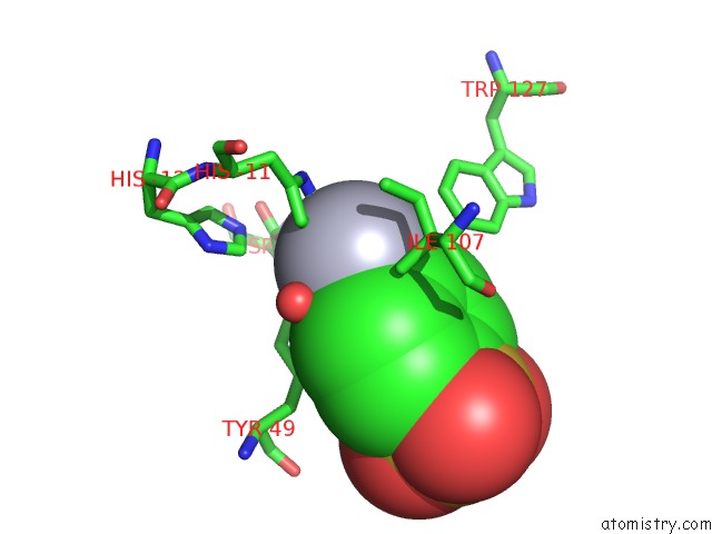
Mono view
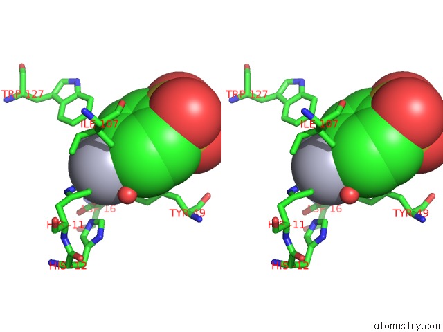
Stereo pair view
Mono view
Stereo pair view
A full contact list of Mercury with other atoms in the Hg binding
site number 1 of Mycobacterium Tuberculosis Kinase Inhibitor Homolog RV0577 within 5.0Å range:
|
Reference:
G.W.Buchko,
N.Echols,
E.M.Flynn,
H.L.Ng,
S.Stephenson,
H.B.Kim,
P.J.Myler,
T.C.Terwilliger,
T.Alber,
C.Y.Kim.
Structural and Biophysical Characterization of the Mycobacterium Tuberculosis Protein RV0577, A Protein Associated with Neutral Red Staining of Virulent Tuberculosis Strains and Homologue of the Streptomyces Coelicolor Protein Kbpa. Biochemistry 2017.
ISSN: ISSN 1520-4995
PubMed: 28692281
DOI: 10.1021/ACS.BIOCHEM.7B00511
Page generated: Fri Aug 8 10:14:38 2025
ISSN: ISSN 1520-4995
PubMed: 28692281
DOI: 10.1021/ACS.BIOCHEM.7B00511
Last articles
Mn in 3OBAMn in 3OB8
Mn in 3O3H
Mn in 3OCH
Mn in 3NIO
Mn in 3NWK
Mn in 3O1R
Mn in 3NVT
Mn in 3O1P
Mn in 3O1O