Mercury »
PDB 3kbc-3wa8 »
3vus »
Mercury in PDB 3vus: Escherichia Coli Pgab N-Terminal Domain
Protein crystallography data
The structure of Escherichia Coli Pgab N-Terminal Domain, PDB code: 3vus
was solved by
T.Nishiyama,
H.Noguchi,
H.Yoshida,
S.-Y.Park,
J.R.H.Tame,
with X-Ray Crystallography technique. A brief refinement statistics is given in the table below:
| Resolution Low / High (Å) | 25.00 / 1.65 |
| Space group | P 1 21 1 |
| Cell size a, b, c (Å), α, β, γ (°) | 39.559, 53.114, 144.170, 90.00, 95.25, 90.00 |
| R / Rfree (%) | 20.5 / 25.6 |
Other elements in 3vus:
The structure of Escherichia Coli Pgab N-Terminal Domain also contains other interesting chemical elements:
| Zinc | (Zn) | 2 atoms |
Mercury Binding Sites:
The binding sites of Mercury atom in the Escherichia Coli Pgab N-Terminal Domain
(pdb code 3vus). This binding sites where shown within
5.0 Angstroms radius around Mercury atom.
In total only one binding site of Mercury was determined in the Escherichia Coli Pgab N-Terminal Domain, PDB code: 3vus:
In total only one binding site of Mercury was determined in the Escherichia Coli Pgab N-Terminal Domain, PDB code: 3vus:
Mercury binding site 1 out of 1 in 3vus
Go back to
Mercury binding site 1 out
of 1 in the Escherichia Coli Pgab N-Terminal Domain
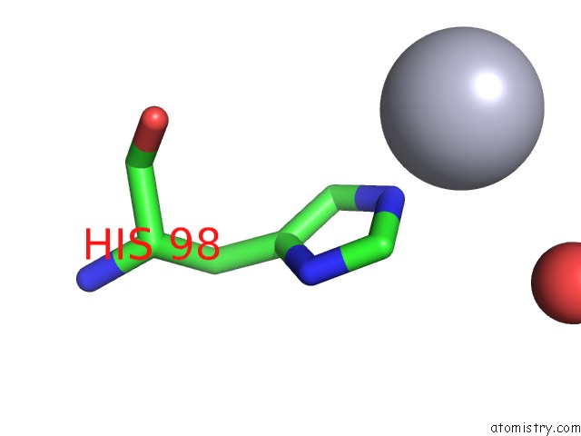
Mono view
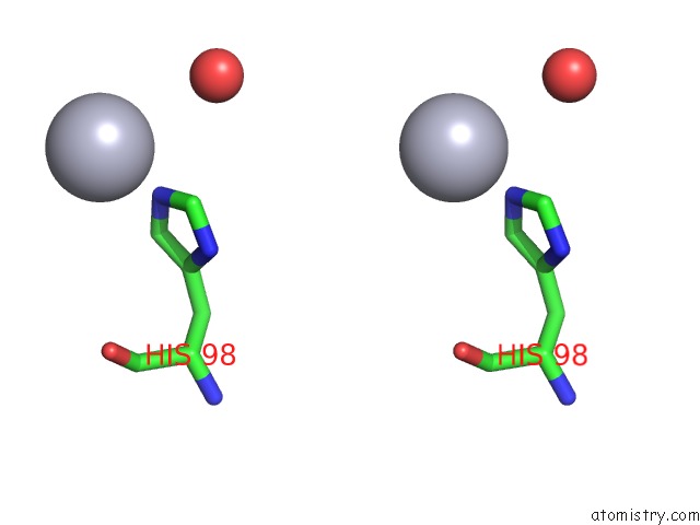
Stereo pair view
Mono view
Stereo pair view
A full contact list of Mercury with other atoms in the Hg binding
site number 1 of Escherichia Coli Pgab N-Terminal Domain within 5.0Å range:
|
Reference:
T.Nishiyama,
H.Noguchi,
H.Yoshida,
S.Y.Park,
J.R.Tame.
The Structure of the Deacetylase Domain of Escherichia Coli Pgab, An Enzyme Required For Biofilm Formation: A Circularly Permuted Member of the Carbohydrate Esterase 4 Family Acta Crystallogr.,Sect.D V. 69 44 2013.
ISSN: ISSN 0907-4449
PubMed: 23275162
DOI: 10.1107/S0907444912042059
Page generated: Fri Aug 8 10:16:40 2025
ISSN: ISSN 0907-4449
PubMed: 23275162
DOI: 10.1107/S0907444912042059
Last articles
Mg in 9MO5Mg in 9MO4
Mg in 9MMI
Mg in 9MM6
Mg in 9MM2
Mg in 9MHS
Mg in 9MJ5
Mg in 9MHG
Mg in 9MHH
Mg in 9MHF