Mercury »
PDB 4ihd-4q7w »
4k7m »
Mercury in PDB 4k7m: Crystal Structure of Rnase S Variant (K7C/Q11C) with Bound Mercury Ions
Enzymatic activity of Crystal Structure of Rnase S Variant (K7C/Q11C) with Bound Mercury Ions
All present enzymatic activity of Crystal Structure of Rnase S Variant (K7C/Q11C) with Bound Mercury Ions:
3.1.27.5;
3.1.27.5;
Protein crystallography data
The structure of Crystal Structure of Rnase S Variant (K7C/Q11C) with Bound Mercury Ions, PDB code: 4k7m
was solved by
M.Genz,
N.Straeter,
with X-Ray Crystallography technique. A brief refinement statistics is given in the table below:
| Resolution Low / High (Å) | 21.93 / 1.80 |
| Space group | P 31 2 1 |
| Cell size a, b, c (Å), α, β, γ (°) | 43.870, 43.870, 96.361, 90.00, 90.00, 120.00 |
| R / Rfree (%) | 16.5 / 20.5 |
Mercury Binding Sites:
The binding sites of Mercury atom in the Crystal Structure of Rnase S Variant (K7C/Q11C) with Bound Mercury Ions
(pdb code 4k7m). This binding sites where shown within
5.0 Angstroms radius around Mercury atom.
In total 2 binding sites of Mercury where determined in the Crystal Structure of Rnase S Variant (K7C/Q11C) with Bound Mercury Ions, PDB code: 4k7m:
Jump to Mercury binding site number: 1; 2;
In total 2 binding sites of Mercury where determined in the Crystal Structure of Rnase S Variant (K7C/Q11C) with Bound Mercury Ions, PDB code: 4k7m:
Jump to Mercury binding site number: 1; 2;
Mercury binding site 1 out of 2 in 4k7m
Go back to
Mercury binding site 1 out
of 2 in the Crystal Structure of Rnase S Variant (K7C/Q11C) with Bound Mercury Ions
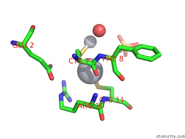
Mono view
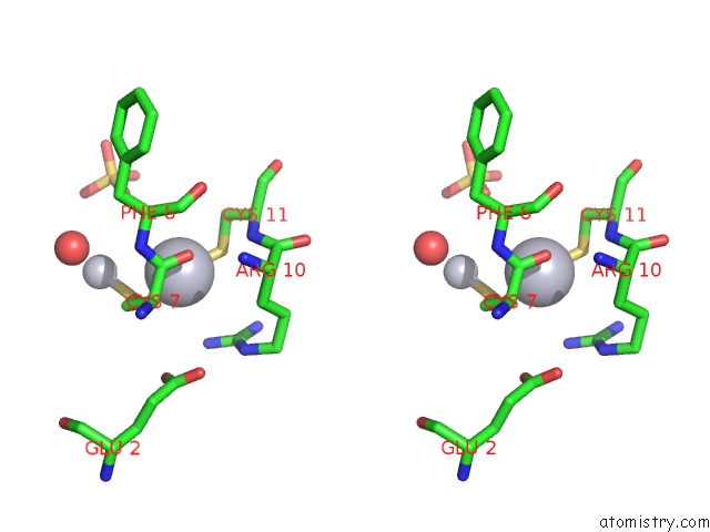
Stereo pair view
Mono view
Stereo pair view
A full contact list of Mercury with other atoms in the Hg binding
site number 1 of Crystal Structure of Rnase S Variant (K7C/Q11C) with Bound Mercury Ions within 5.0Å range:
|
Mercury binding site 2 out of 2 in 4k7m
Go back to
Mercury binding site 2 out
of 2 in the Crystal Structure of Rnase S Variant (K7C/Q11C) with Bound Mercury Ions
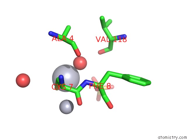
Mono view
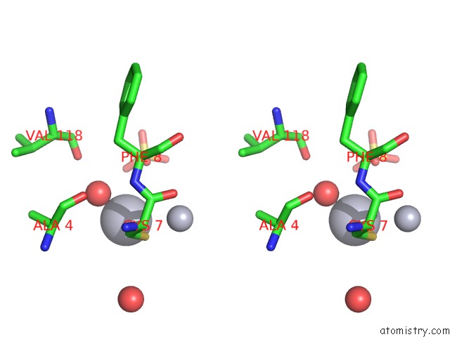
Stereo pair view
Mono view
Stereo pair view
A full contact list of Mercury with other atoms in the Hg binding
site number 2 of Crystal Structure of Rnase S Variant (K7C/Q11C) with Bound Mercury Ions within 5.0Å range:
|
Reference:
M.Genz,
D.Singer,
E.Hey-Hawkins,
R.Hoffmann,
N.Straeter.
Crystal Structure of Apo- and Metalated Thiolate Containing Rnase S As Structural Basis For the Design of Artificial Metalloenzymes By Peptide- Protein Complementation Z.Anorg.Allg.Chem. V. 639 2395 2013.
DOI: 10.1002/ZAAC.201300410
Page generated: Fri Aug 8 10:29:29 2025
DOI: 10.1002/ZAAC.201300410
Last articles
Mo in 4ZOHMo in 4XPI
Mo in 4WZB
Mo in 4WZA
Mo in 4WNA
Mo in 4WN9
Mo in 4WES
Mo in 4USA
Mo in 4US9
Mo in 4UHX