Mercury »
PDB 4ihd-4q7w »
4m7r »
Mercury in PDB 4m7r: Crystal Structure of the N-Terminal Methyltransferase-Like Domain of Anamorsin
Protein crystallography data
The structure of Crystal Structure of the N-Terminal Methyltransferase-Like Domain of Anamorsin, PDB code: 4m7r
was solved by
G.Song,
Z.-J.Liu,
with X-Ray Crystallography technique. A brief refinement statistics is given in the table below:
| Resolution Low / High (Å) | 30.43 / 1.80 |
| Space group | P 61 |
| Cell size a, b, c (Å), α, β, γ (°) | 79.508, 79.508, 101.750, 90.00, 90.00, 120.00 |
| R / Rfree (%) | 18.7 / 22.6 |
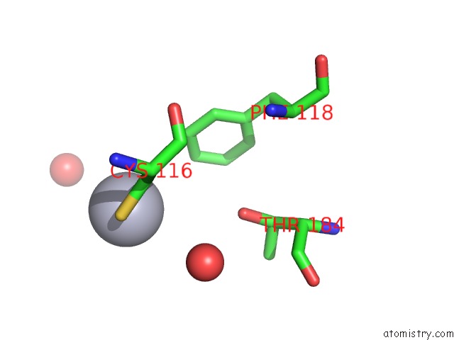
Mercury Binding Sites:
The binding sites of Mercury atom in the Crystal Structure of the N-Terminal Methyltransferase-Like Domain of Anamorsin
(pdb code 4m7r). This binding sites where shown within
5.0 Angstroms radius around Mercury atom.
In total only one binding site of Mercury was determined in the Crystal Structure of the N-Terminal Methyltransferase-Like Domain of Anamorsin, PDB code: 4m7r:
In total only one binding site of Mercury was determined in the Crystal Structure of the N-Terminal Methyltransferase-Like Domain of Anamorsin, PDB code: 4m7r:
Mercury binding site 1 out of 1 in 4m7r
Go back to
Mercury binding site 1 out
of 1 in the Crystal Structure of the N-Terminal Methyltransferase-Like Domain of Anamorsin
Mono view
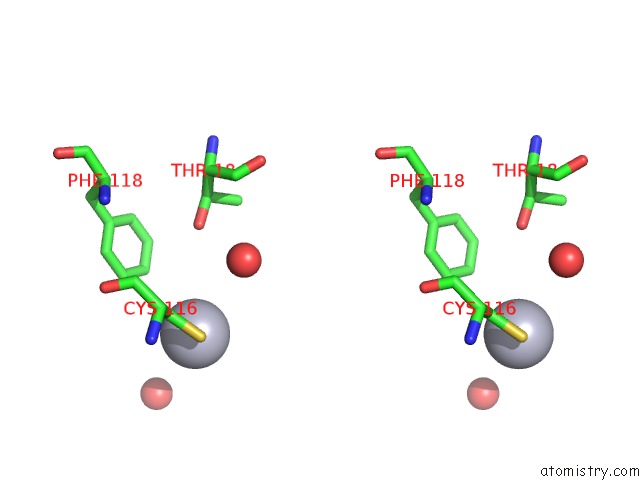
Stereo pair view
Mono view
Stereo pair view
A full contact list of Mercury with other atoms in the Hg binding
site number 1 of Crystal Structure of the N-Terminal Methyltransferase-Like Domain of Anamorsin within 5.0Å range:
|
Reference:
G.Song,
C.Cheng,
Y.Li,
N.Shaw,
Z.Xiao,
Z.-J.Liu.
Crystal Structure of the N-Terminal Methyltransferase-Like Domain of Anamorsin Proteins 2013.
ISSN: ESSN 1097-0134
PubMed: 24123282
DOI: 10.1002/PROT.24443
Page generated: Sun Aug 11 04:57:25 2024
ISSN: ESSN 1097-0134
PubMed: 24123282
DOI: 10.1002/PROT.24443
Last articles
Zn in 9MJ5Zn in 9HNW
Zn in 9G0L
Zn in 9FNE
Zn in 9DZN
Zn in 9E0I
Zn in 9D32
Zn in 9DAK
Zn in 8ZXC
Zn in 8ZUF