Mercury »
PDB 5lf8-6bvg »
5oon »
Mercury in PDB 5oon: Structure of Undecaprenyl-Pyrophosphate Phosphatase, Baca
Enzymatic activity of Structure of Undecaprenyl-Pyrophosphate Phosphatase, Baca
All present enzymatic activity of Structure of Undecaprenyl-Pyrophosphate Phosphatase, Baca:
3.6.1.27;
3.6.1.27;
Protein crystallography data
The structure of Structure of Undecaprenyl-Pyrophosphate Phosphatase, Baca, PDB code: 5oon
was solved by
C.-Y.Huang,
V.Olieric,
R.Warshamanage,
M.Wang,
N.Howe,
M.E.I.Ghachi,
D.Weichert,
F.Kerff,
P.Stansfeld,
T.Touze,
M.Caffrey,
with X-Ray Crystallography technique. A brief refinement statistics is given in the table below:
| Resolution Low / High (Å) | 44.46 / 2.60 |
| Space group | C 2 2 2 |
| Cell size a, b, c (Å), α, β, γ (°) | 113.260, 145.000, 40.490, 90.00, 90.00, 90.00 |
| R / Rfree (%) | 20.6 / 24.4 |
Mercury Binding Sites:
The binding sites of Mercury atom in the Structure of Undecaprenyl-Pyrophosphate Phosphatase, Baca
(pdb code 5oon). This binding sites where shown within
5.0 Angstroms radius around Mercury atom.
In total 2 binding sites of Mercury where determined in the Structure of Undecaprenyl-Pyrophosphate Phosphatase, Baca, PDB code: 5oon:
Jump to Mercury binding site number: 1; 2;
In total 2 binding sites of Mercury where determined in the Structure of Undecaprenyl-Pyrophosphate Phosphatase, Baca, PDB code: 5oon:
Jump to Mercury binding site number: 1; 2;
Mercury binding site 1 out of 2 in 5oon
Go back to
Mercury binding site 1 out
of 2 in the Structure of Undecaprenyl-Pyrophosphate Phosphatase, Baca
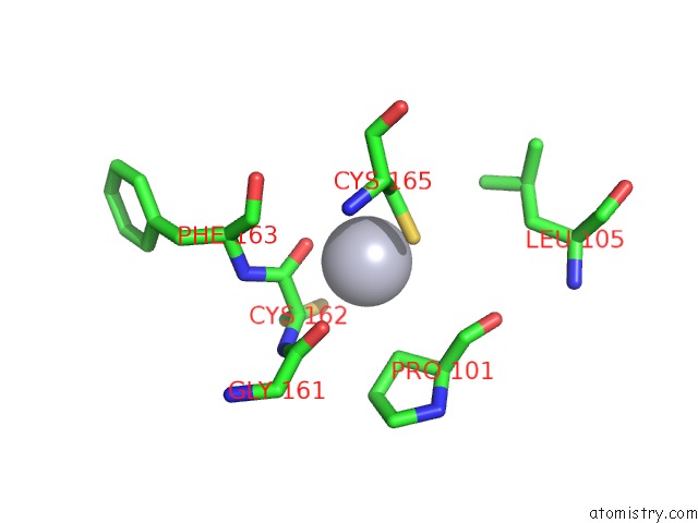
Mono view
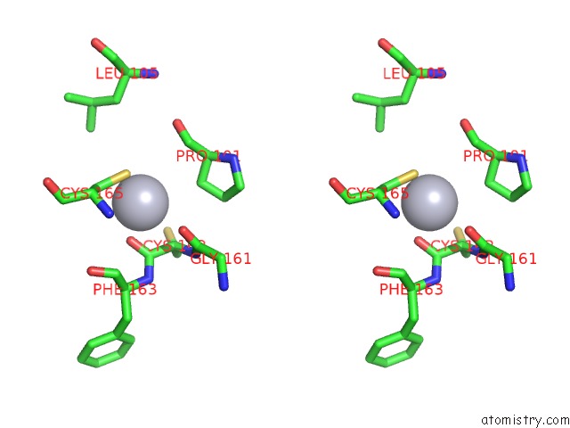
Stereo pair view
Mono view
Stereo pair view
A full contact list of Mercury with other atoms in the Hg binding
site number 1 of Structure of Undecaprenyl-Pyrophosphate Phosphatase, Baca within 5.0Å range:
|
Mercury binding site 2 out of 2 in 5oon
Go back to
Mercury binding site 2 out
of 2 in the Structure of Undecaprenyl-Pyrophosphate Phosphatase, Baca
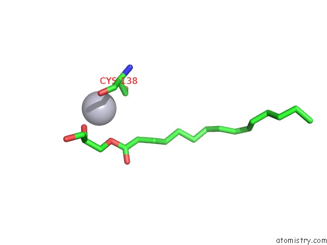
Mono view
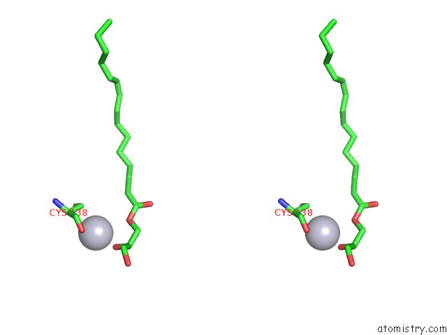
Stereo pair view
Mono view
Stereo pair view
A full contact list of Mercury with other atoms in the Hg binding
site number 2 of Structure of Undecaprenyl-Pyrophosphate Phosphatase, Baca within 5.0Å range:
|
Reference:
M.El Ghachi,
N.Howe,
C.Y.Huang,
V.Olieric,
R.Warshamanage,
T.Touze,
D.Weichert,
P.J.Stansfeld,
M.Wang,
F.Kerff,
M.Caffrey.
Crystal Structure of Undecaprenyl-Pyrophosphate Phosphatase and Its Role in Peptidoglycan Biosynthesis. Nat Commun V. 9 1078 2018.
ISSN: ESSN 2041-1723
PubMed: 29540682
DOI: 10.1038/S41467-018-03477-5
Page generated: Fri Aug 8 10:57:35 2025
ISSN: ESSN 2041-1723
PubMed: 29540682
DOI: 10.1038/S41467-018-03477-5
Last articles
I in 5W1HI in 5W1I
I in 5W0M
I in 5W0N
I in 5W0B
I in 5W0J
I in 5V65
I in 5VTE
I in 5VF1
I in 5VQ5