Mercury »
PDB 5lf8-6bvg »
5tiq »
Mercury in PDB 5tiq: The Structure of the Major Capsid Protein of Pbcv-1
Protein crystallography data
The structure of The Structure of the Major Capsid Protein of Pbcv-1, PDB code: 5tiq
was solved by
T.Klose,
C.De Castro,
I.Speciale,
A.Molinaro,
J.L.Van Etten,
M.G.Rossmann,
with X-Ray Crystallography technique. A brief refinement statistics is given in the table below:
| Resolution Low / High (Å) | 43.31 / 2.54 |
| Space group | P 41 3 2 |
| Cell size a, b, c (Å), α, β, γ (°) | 188.763, 188.763, 188.763, 90.00, 90.00, 90.00 |
| R / Rfree (%) | 17.9 / 22.3 |
Mercury Binding Sites:
The binding sites of Mercury atom in the The Structure of the Major Capsid Protein of Pbcv-1
(pdb code 5tiq). This binding sites where shown within
5.0 Angstroms radius around Mercury atom.
In total 4 binding sites of Mercury where determined in the The Structure of the Major Capsid Protein of Pbcv-1, PDB code: 5tiq:
Jump to Mercury binding site number: 1; 2; 3; 4;
In total 4 binding sites of Mercury where determined in the The Structure of the Major Capsid Protein of Pbcv-1, PDB code: 5tiq:
Jump to Mercury binding site number: 1; 2; 3; 4;
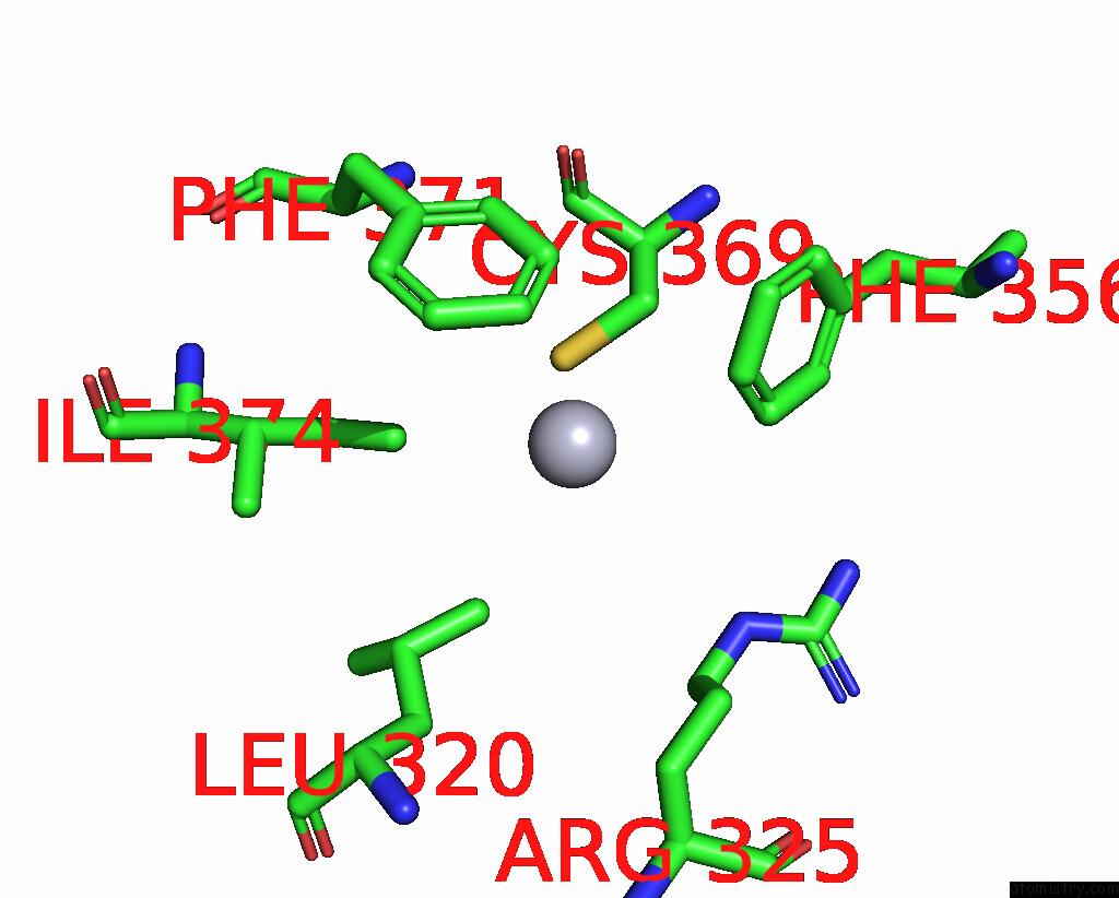
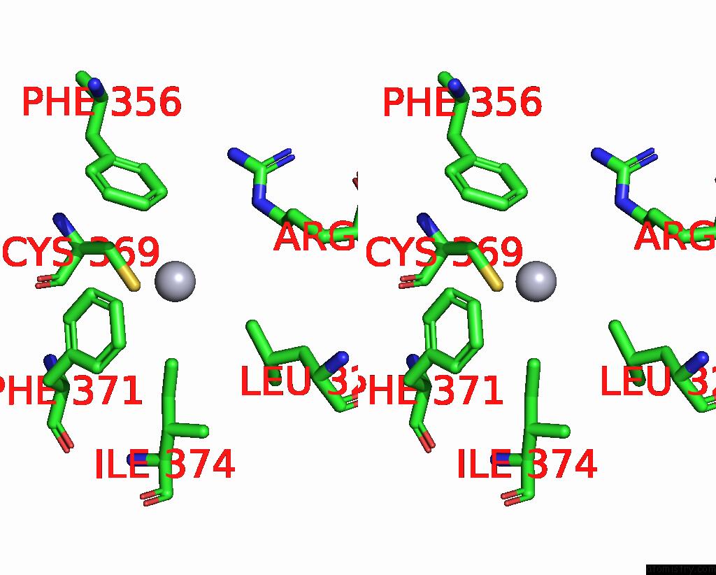
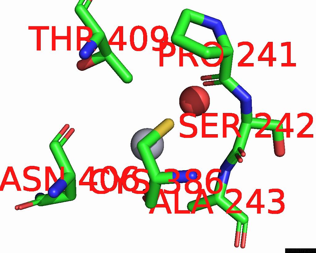
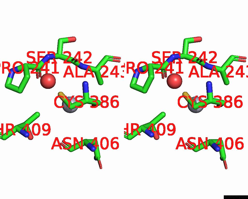
Mercury binding site 1 out of 4 in 5tiq
Go back to
Mercury binding site 1 out
of 4 in the The Structure of the Major Capsid Protein of Pbcv-1
Mono view
Stereo pair view
Mono view
Stereo pair view
A full contact list of Mercury with other atoms in the Hg binding
site number 1 of The Structure of the Major Capsid Protein of Pbcv-1 within 5.0Å range:
|
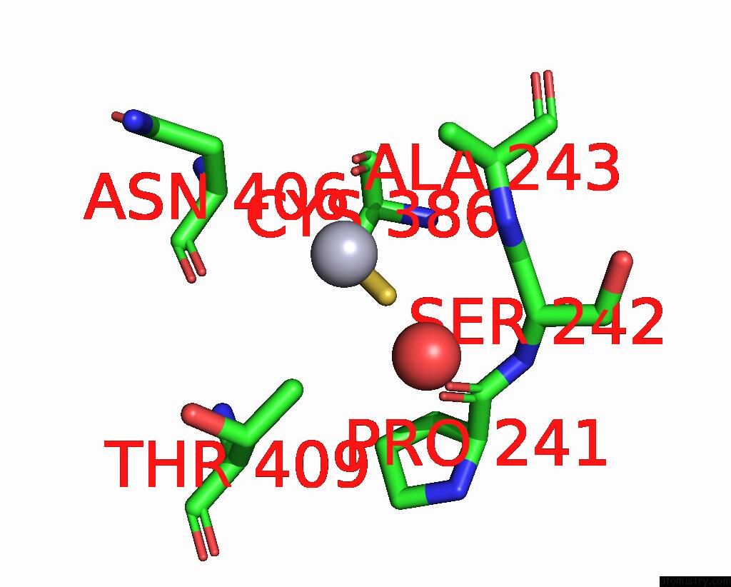
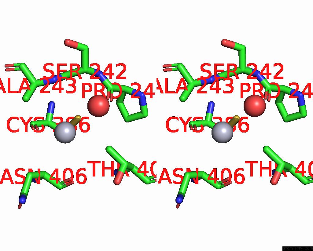
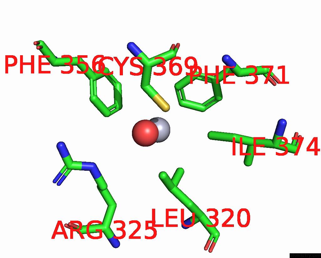
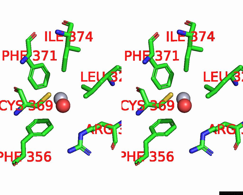
Mercury binding site 2 out of 4 in 5tiq
Go back to
Mercury binding site 2 out
of 4 in the The Structure of the Major Capsid Protein of Pbcv-1
Mono view
Stereo pair view
Mono view
Stereo pair view
A full contact list of Mercury with other atoms in the Hg binding
site number 2 of The Structure of the Major Capsid Protein of Pbcv-1 within 5.0Å range:
|
Mercury binding site 3 out of 4 in 5tiq
Go back to
Mercury binding site 3 out
of 4 in the The Structure of the Major Capsid Protein of Pbcv-1
Mono view
Stereo pair view
Mono view
Stereo pair view
A full contact list of Mercury with other atoms in the Hg binding
site number 3 of The Structure of the Major Capsid Protein of Pbcv-1 within 5.0Å range:
|
Mercury binding site 4 out of 4 in 5tiq
Go back to
Mercury binding site 4 out
of 4 in the The Structure of the Major Capsid Protein of Pbcv-1
Mono view
Stereo pair view
Mono view
Stereo pair view
A full contact list of Mercury with other atoms in the Hg binding
site number 4 of The Structure of the Major Capsid Protein of Pbcv-1 within 5.0Å range:
|
Reference:
C.De Castro,
T.Klose,
I.Speciale,
R.Lanzetta,
A.Molinaro,
J.L.Van Etten,
M.G.Rossmann.
Structure of the Chlorovirus Pbcv-1 Major Capsid Glycoprotein Determined By Combining Crystallographic and Carbohydrate Molecular Modeling Approaches. Proc. Natl. Acad. Sci. V. 115 E44 2018U.S.A..
ISSN: ESSN 1091-6490
PubMed: 29255015
DOI: 10.1073/PNAS.1613432115
Page generated: Fri Aug 8 10:59:27 2025
ISSN: ESSN 1091-6490
PubMed: 29255015
DOI: 10.1073/PNAS.1613432115
Last articles
K in 1PQJK in 1PCQ
K in 1PQI
K in 1PQD
K in 1PKN
K in 1PI5
K in 1PI4
K in 1P9E
K in 1P7L
K in 1P7B