Mercury »
PDB 5lf8-6bvg »
5w2k »
Mercury in PDB 5w2k: Crystal Structure of Mutant Cj Ycei Protein (Cj-G34C) with Hydroxymercuribenzoic Acid Guest Structure
Protein crystallography data
The structure of Crystal Structure of Mutant Cj Ycei Protein (Cj-G34C) with Hydroxymercuribenzoic Acid Guest Structure, PDB code: 5w2k
was solved by
T.R.Huber,
C.D.Snow,
with X-Ray Crystallography technique. A brief refinement statistics is given in the table below:
| Resolution Low / High (Å) | 38.71 / 2.78 |
| Space group | P 6 2 2 |
| Cell size a, b, c (Å), α, β, γ (°) | 178.812, 178.812, 50.640, 90.00, 90.00, 120.00 |
| R / Rfree (%) | 22.1 / 26.1 |
Mercury Binding Sites:
The binding sites of Mercury atom in the Crystal Structure of Mutant Cj Ycei Protein (Cj-G34C) with Hydroxymercuribenzoic Acid Guest Structure
(pdb code 5w2k). This binding sites where shown within
5.0 Angstroms radius around Mercury atom.
In total only one binding site of Mercury was determined in the Crystal Structure of Mutant Cj Ycei Protein (Cj-G34C) with Hydroxymercuribenzoic Acid Guest Structure, PDB code: 5w2k:
In total only one binding site of Mercury was determined in the Crystal Structure of Mutant Cj Ycei Protein (Cj-G34C) with Hydroxymercuribenzoic Acid Guest Structure, PDB code: 5w2k:
Mercury binding site 1 out of 1 in 5w2k
Go back to
Mercury binding site 1 out
of 1 in the Crystal Structure of Mutant Cj Ycei Protein (Cj-G34C) with Hydroxymercuribenzoic Acid Guest Structure
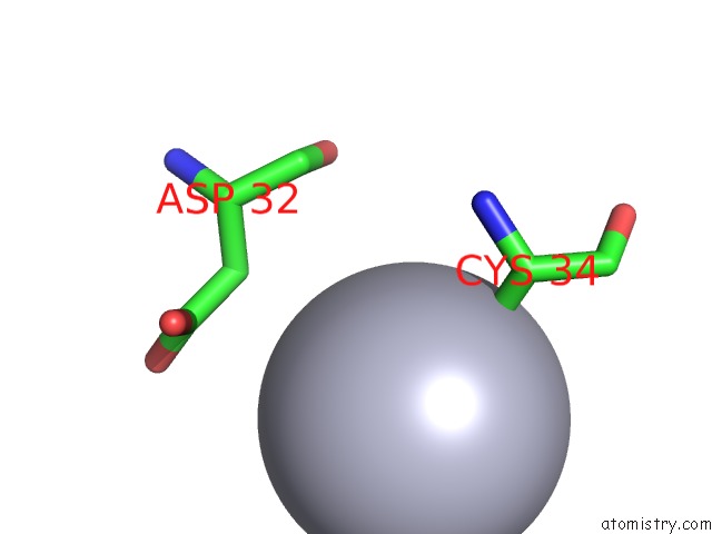
Mono view
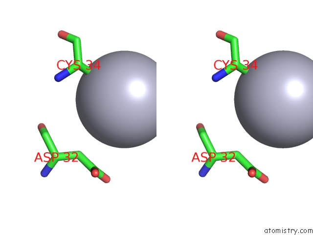
Stereo pair view
Mono view
Stereo pair view
A full contact list of Mercury with other atoms in the Hg binding
site number 1 of Crystal Structure of Mutant Cj Ycei Protein (Cj-G34C) with Hydroxymercuribenzoic Acid Guest Structure within 5.0Å range:
|
Reference:
T.R.Huber,
E.C.Mcpherson,
C.E.Keating,
C.D.Snow.
Installing Guest Molecules at Specific Sites Within Scaffold Protein Crystals. Bioconjug. Chem. V. 29 17 2018.
ISSN: ISSN 1520-4812
PubMed: 29232505
DOI: 10.1021/ACS.BIOCONJCHEM.7B00668
Page generated: Fri Aug 8 11:00:21 2025
ISSN: ISSN 1520-4812
PubMed: 29232505
DOI: 10.1021/ACS.BIOCONJCHEM.7B00668
Last articles
I in 4OHMI in 4OHK
I in 4OEK
I in 4OEW
I in 4OC5
I in 4OC4
I in 4OC3
I in 4OC1
I in 4OC2
I in 4NHB