Mercury »
PDB 6rkn-7byf »
6sfq »
Mercury in PDB 6sfq: Atomic Resolution Structure of Human Carbonic Anhydrase II in Complex with (R)-5-Phenyloxazolidine-2,4-Dione
Enzymatic activity of Atomic Resolution Structure of Human Carbonic Anhydrase II in Complex with (R)-5-Phenyloxazolidine-2,4-Dione
All present enzymatic activity of Atomic Resolution Structure of Human Carbonic Anhydrase II in Complex with (R)-5-Phenyloxazolidine-2,4-Dione:
4.2.1.1;
4.2.1.1;
Protein crystallography data
The structure of Atomic Resolution Structure of Human Carbonic Anhydrase II in Complex with (R)-5-Phenyloxazolidine-2,4-Dione, PDB code: 6sfq
was solved by
S.Gloeckner,
K.Ngo,
A.Heine,
G.Klebe,
with X-Ray Crystallography technique. A brief refinement statistics is given in the table below:
| Resolution Low / High (Å) | 41.06 / 1.00 |
| Space group | P 1 21 1 |
| Cell size a, b, c (Å), α, β, γ (°) | 42.410, 41.608, 72.135, 90.00, 104.48, 90.00 |
| R / Rfree (%) | 12.6 / 14.7 |
Other elements in 6sfq:
The structure of Atomic Resolution Structure of Human Carbonic Anhydrase II in Complex with (R)-5-Phenyloxazolidine-2,4-Dione also contains other interesting chemical elements:
| Zinc | (Zn) | 1 atom |
| Sodium | (Na) | 1 atom |
Mercury Binding Sites:
The binding sites of Mercury atom in the Atomic Resolution Structure of Human Carbonic Anhydrase II in Complex with (R)-5-Phenyloxazolidine-2,4-Dione
(pdb code 6sfq). This binding sites where shown within
5.0 Angstroms radius around Mercury atom.
In total 3 binding sites of Mercury where determined in the Atomic Resolution Structure of Human Carbonic Anhydrase II in Complex with (R)-5-Phenyloxazolidine-2,4-Dione, PDB code: 6sfq:
Jump to Mercury binding site number: 1; 2; 3;
In total 3 binding sites of Mercury where determined in the Atomic Resolution Structure of Human Carbonic Anhydrase II in Complex with (R)-5-Phenyloxazolidine-2,4-Dione, PDB code: 6sfq:
Jump to Mercury binding site number: 1; 2; 3;
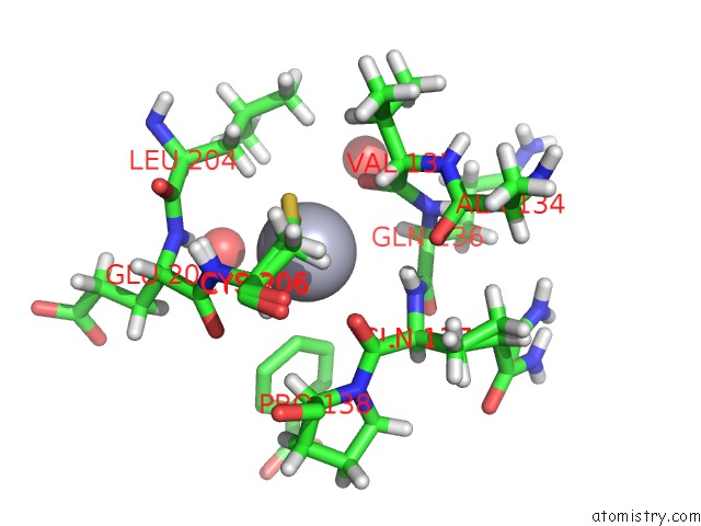
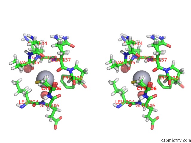
Mercury binding site 1 out of 3 in 6sfq
Go back to
Mercury binding site 1 out
of 3 in the Atomic Resolution Structure of Human Carbonic Anhydrase II in Complex with (R)-5-Phenyloxazolidine-2,4-Dione
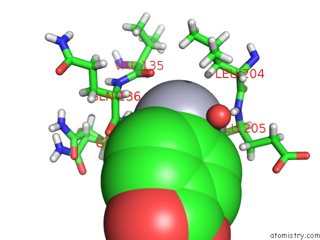
Mono view
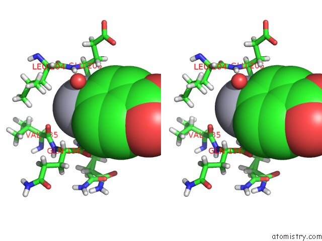
Stereo pair view
Mono view
Stereo pair view
A full contact list of Mercury with other atoms in the Hg binding
site number 1 of Atomic Resolution Structure of Human Carbonic Anhydrase II in Complex with (R)-5-Phenyloxazolidine-2,4-Dione within 5.0Å range:
|
Mercury binding site 2 out of 3 in 6sfq
Go back to
Mercury binding site 2 out
of 3 in the Atomic Resolution Structure of Human Carbonic Anhydrase II in Complex with (R)-5-Phenyloxazolidine-2,4-Dione
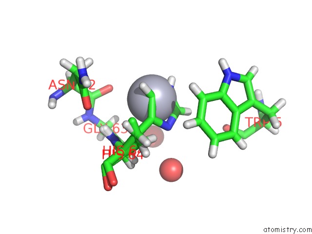
Mono view
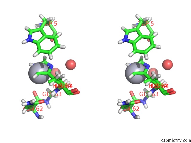
Stereo pair view
Mono view
Stereo pair view
A full contact list of Mercury with other atoms in the Hg binding
site number 2 of Atomic Resolution Structure of Human Carbonic Anhydrase II in Complex with (R)-5-Phenyloxazolidine-2,4-Dione within 5.0Å range:
|
Mercury binding site 3 out of 3 in 6sfq
Go back to
Mercury binding site 3 out
of 3 in the Atomic Resolution Structure of Human Carbonic Anhydrase II in Complex with (R)-5-Phenyloxazolidine-2,4-Dione
Mono view
Stereo pair view
Mono view
Stereo pair view
A full contact list of Mercury with other atoms in the Hg binding
site number 3 of Atomic Resolution Structure of Human Carbonic Anhydrase II in Complex with (R)-5-Phenyloxazolidine-2,4-Dione within 5.0Å range:
|
Reference:
S.Gloeckner,
K.Ngo,
A.Heine,
G.Klebe.
Atomic Resolution Structure of Human Carbonic Anhydrase II in Complex with (R)-5-Phenyloxazolidine-2,4-Dione To Be Published.
Page generated: Fri Aug 8 11:22:52 2025
Last articles
Na in 3I3CNa in 3I31
Na in 3I2W
Na in 3I1J
Na in 3I04
Na in 3I01
Na in 3I0X
Na in 3I0W
Na in 3HWX
Na in 3HZN