Mercury »
PDB 8phl-9fei »
8q0c »
Mercury in PDB 8q0c: Human Carbonic Anhydrase II Containing 3-Fluorotyrosine
Enzymatic activity of Human Carbonic Anhydrase II Containing 3-Fluorotyrosine
All present enzymatic activity of Human Carbonic Anhydrase II Containing 3-Fluorotyrosine:
4.2.1.1; 4.2.1.69;
4.2.1.1; 4.2.1.69;
Protein crystallography data
The structure of Human Carbonic Anhydrase II Containing 3-Fluorotyrosine, PDB code: 8q0c
was solved by
L.B.T.Pham,
A.Costantino,
L.Barbieri,
V.Calderone,
E.Luchinat,
L.Banci,
with X-Ray Crystallography technique. A brief refinement statistics is given in the table below:
| Resolution Low / High (Å) | 23.01 / 1.30 |
| Space group | P 1 21 1 |
| Cell size a, b, c (Å), α, β, γ (°) | 41.99, 41.14, 71.21, 90, 104.2, 90 |
| R / Rfree (%) | 16.5 / 19.7 |
Other elements in 8q0c:
The structure of Human Carbonic Anhydrase II Containing 3-Fluorotyrosine also contains other interesting chemical elements:
| Fluorine | (F) | 11 atoms |
| Zinc | (Zn) | 1 atom |
Mercury Binding Sites:
The binding sites of Mercury atom in the Human Carbonic Anhydrase II Containing 3-Fluorotyrosine
(pdb code 8q0c). This binding sites where shown within
5.0 Angstroms radius around Mercury atom.
In total only one binding site of Mercury was determined in the Human Carbonic Anhydrase II Containing 3-Fluorotyrosine, PDB code: 8q0c:
In total only one binding site of Mercury was determined in the Human Carbonic Anhydrase II Containing 3-Fluorotyrosine, PDB code: 8q0c:
Mercury binding site 1 out of 1 in 8q0c
Go back to
Mercury binding site 1 out
of 1 in the Human Carbonic Anhydrase II Containing 3-Fluorotyrosine
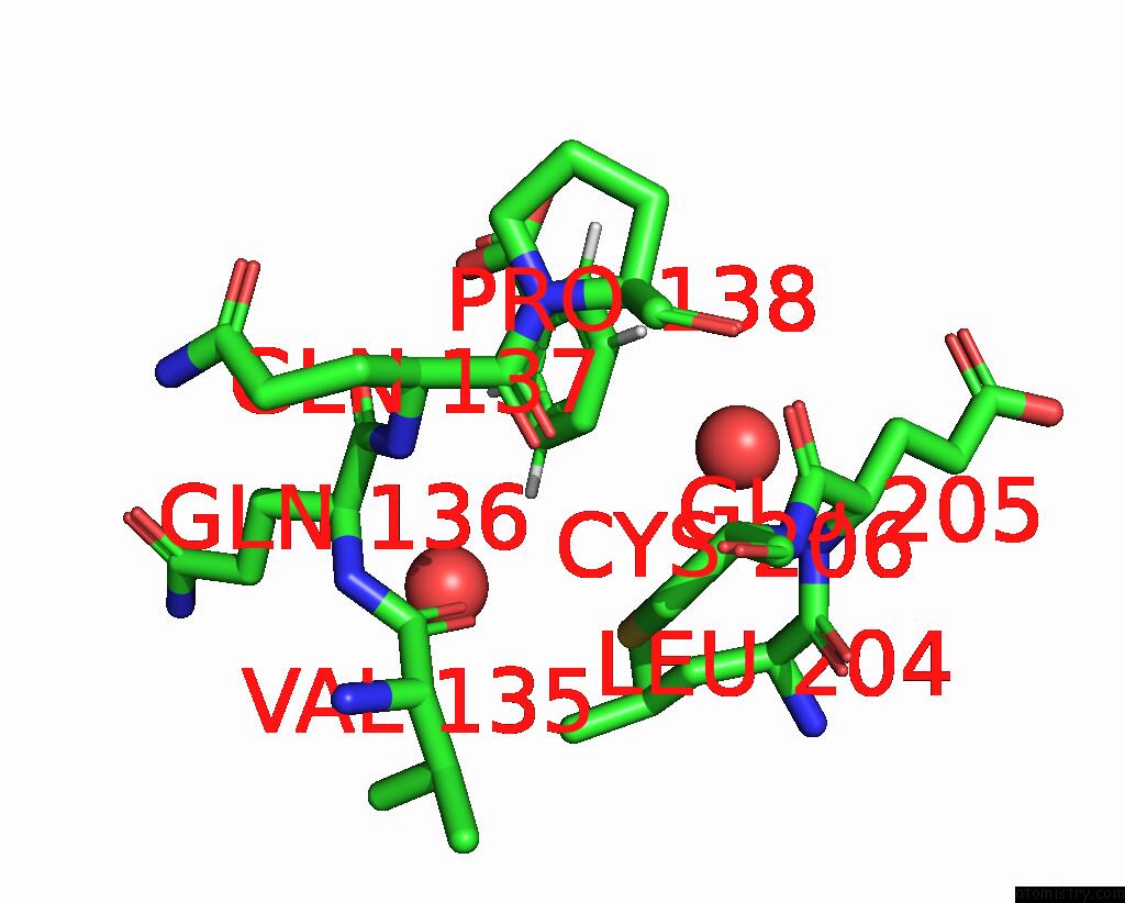
Mono view
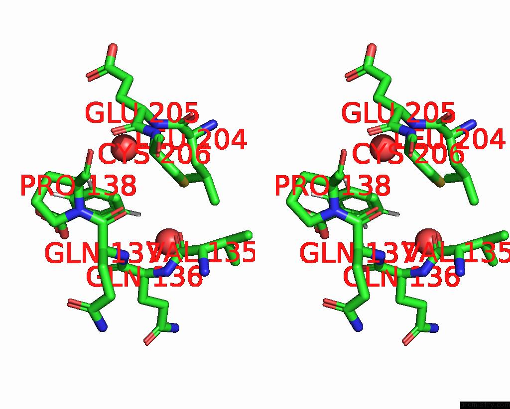
Stereo pair view
Mono view
Stereo pair view
A full contact list of Mercury with other atoms in the Hg binding
site number 1 of Human Carbonic Anhydrase II Containing 3-Fluorotyrosine within 5.0Å range:
|
Reference:
A.Costantino,
L.B.T.Pham,
L.Barbieri,
V.Calderone,
G.Ben-Nissan,
M.Sharon,
L.Banci,
E.Luchinat.
Controlling the Incorporation of Fluorinated Amino Acids in Human Cells and Its Structural Impact. Protein Sci. V. 33 E4910 2024.
ISSN: ESSN 1469-896X
PubMed: 38358125
DOI: 10.1002/PRO.4910
Page generated: Fri Aug 8 11:35:17 2025
ISSN: ESSN 1469-896X
PubMed: 38358125
DOI: 10.1002/PRO.4910
Last articles
Mg in 7TF9Mg in 7TFE
Mg in 7TDP
Mg in 7TFH
Mg in 7TDB
Mg in 7TE5
Mg in 7TDA
Mg in 7TDC
Mg in 7TD8
Mg in 7TD7