Mercury »
PDB 7ca2-7tfw »
7edl »
Mercury in PDB 7edl: Crystal Structure of the Bacterial Ribosomal Decoding Site in Complex with G418 and Hg(II)
Protein crystallography data
The structure of Crystal Structure of the Bacterial Ribosomal Decoding Site in Complex with G418 and Hg(II), PDB code: 7edl
was solved by
J.Kondo,
C.Suzuki,
with X-Ray Crystallography technique. A brief refinement statistics is given in the table below:
| Resolution Low / High (Å) | 48.65 / 2.60 |
| Space group | P 21 21 2 |
| Cell size a, b, c (Å), α, β, γ (°) | 31.64, 97.301, 47.06, 90, 90, 90 |
| R / Rfree (%) | 18.6 / 22.2 |
Mercury Binding Sites:
The binding sites of Mercury atom in the Crystal Structure of the Bacterial Ribosomal Decoding Site in Complex with G418 and Hg(II)
(pdb code 7edl). This binding sites where shown within
5.0 Angstroms radius around Mercury atom.
In total 2 binding sites of Mercury where determined in the Crystal Structure of the Bacterial Ribosomal Decoding Site in Complex with G418 and Hg(II), PDB code: 7edl:
Jump to Mercury binding site number: 1; 2;
In total 2 binding sites of Mercury where determined in the Crystal Structure of the Bacterial Ribosomal Decoding Site in Complex with G418 and Hg(II), PDB code: 7edl:
Jump to Mercury binding site number: 1; 2;
Mercury binding site 1 out of 2 in 7edl
Go back to
Mercury binding site 1 out
of 2 in the Crystal Structure of the Bacterial Ribosomal Decoding Site in Complex with G418 and Hg(II)
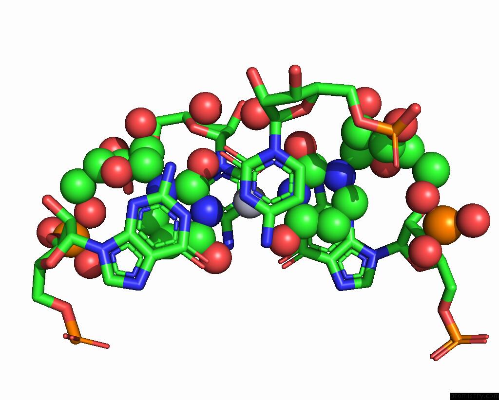
Mono view
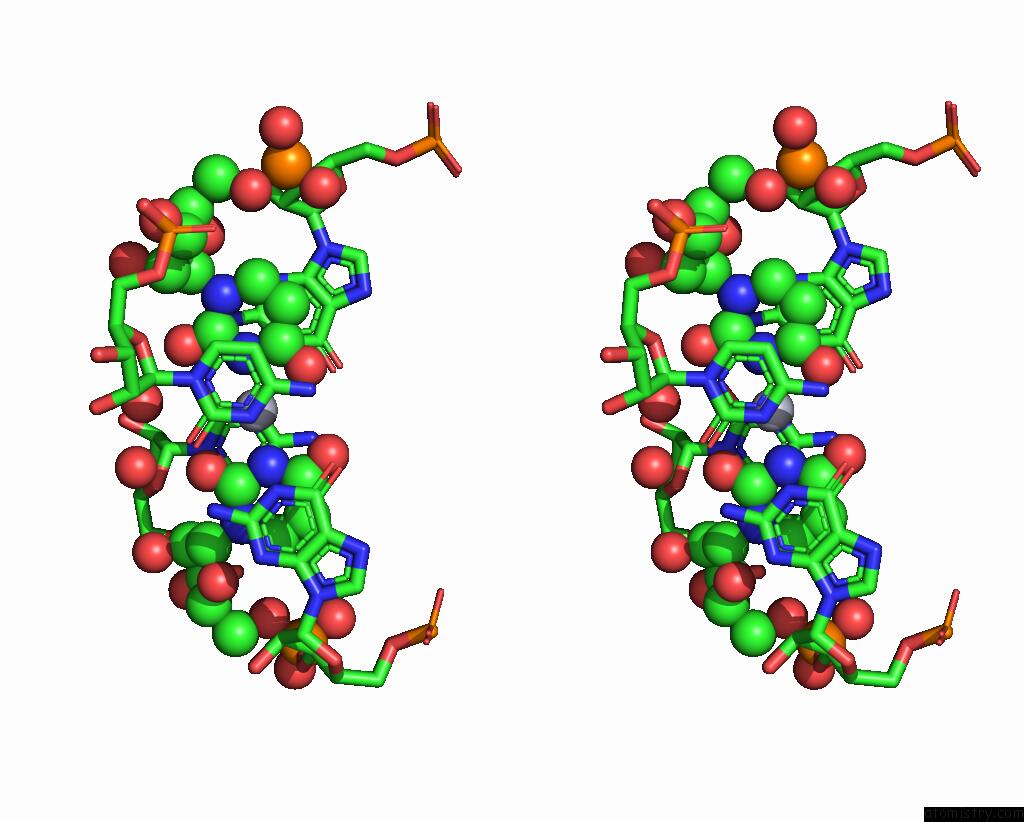
Stereo pair view
Mono view
Stereo pair view
A full contact list of Mercury with other atoms in the Hg binding
site number 1 of Crystal Structure of the Bacterial Ribosomal Decoding Site in Complex with G418 and Hg(II) within 5.0Å range:
|
Mercury binding site 2 out of 2 in 7edl
Go back to
Mercury binding site 2 out
of 2 in the Crystal Structure of the Bacterial Ribosomal Decoding Site in Complex with G418 and Hg(II)
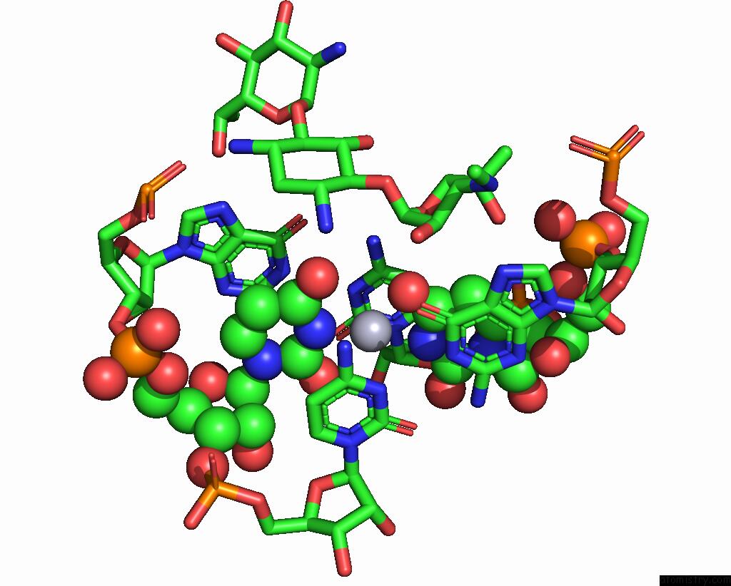
Mono view
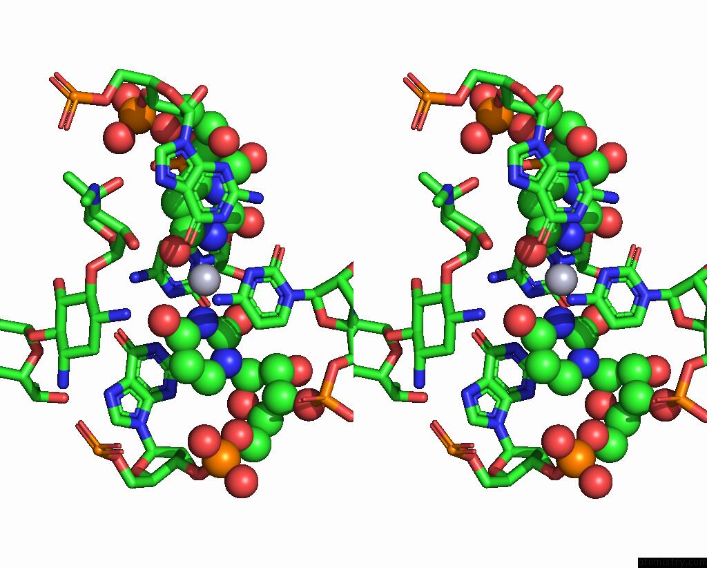
Stereo pair view
Mono view
Stereo pair view
A full contact list of Mercury with other atoms in the Hg binding
site number 2 of Crystal Structure of the Bacterial Ribosomal Decoding Site in Complex with G418 and Hg(II) within 5.0Å range:
|
Reference:
J.Kondo,
C.Suzuki.
Crystal Structure of the Prokaryotic Ribosomal Decoding Site in Complex with G418 and Hg(II) To Be Published.
Page generated: Fri Aug 8 11:27:12 2025
Last articles
K in 3SBQK in 3S83
K in 3S28
K in 3S29
K in 3S27
K in 3S7K
K in 3S5O
K in 3S5N
K in 3S49
K in 3S3X